. 2011 Jan 7;108(1):51-9.
doi: 10.1161/CIRCRESAHA.110.233262. Epub 2010 Nov 24.

A dynamic notch injury response activates epicardium and contributes to fibrosis repair

Affiliations

A dynamic notch injury response activates epicardium and contributes to fibrosis repair

Jamie L Russell et al. Circ Res. .

Abstract

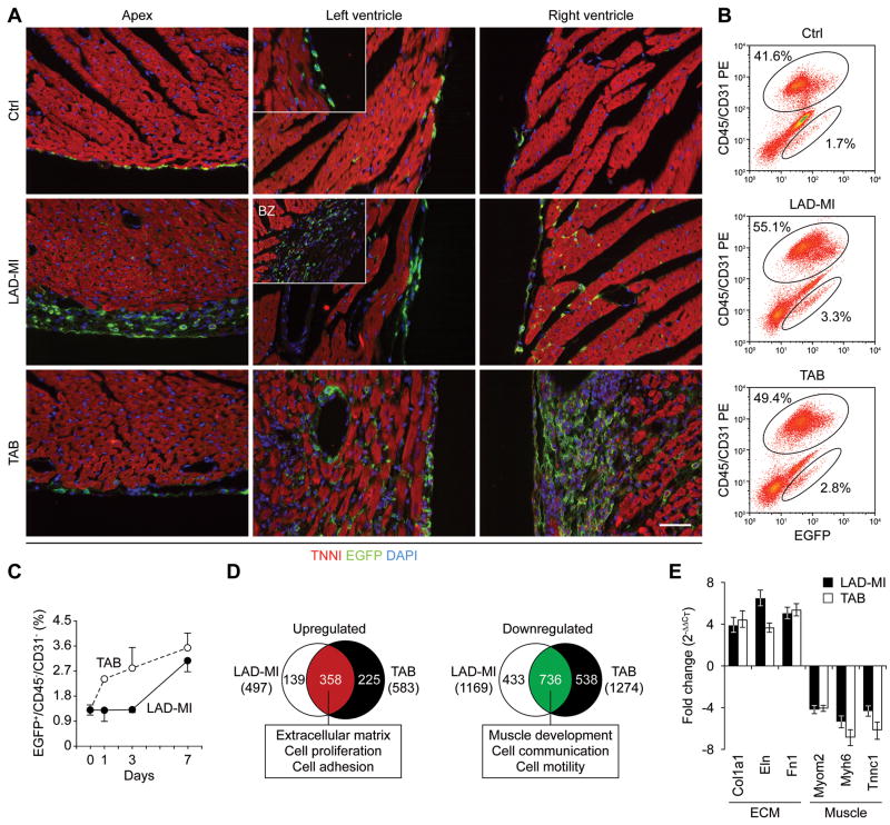
Rationale: Transgenic Notch reporter mice express enhanced green fluorescent protein in cells with C-promoter binding factor-1 response element transcriptional activity (CBF1-RE(x)₄-EGFP), providing a unique and powerful tool for identifying and isolating "Notch-activated" progenitors.

Objective: We asked whether, as in other tissues of this mouse, EGFP localized and functionally tagged adult cardiac tissue progenitors, and, if so, whether this cell-based signal could serve as a quantitative and qualitative biosensor of the injury repair response of the heart.

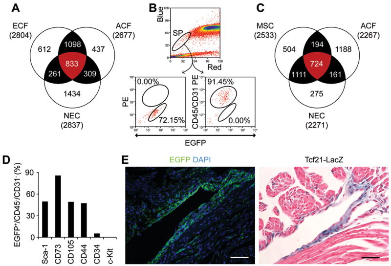
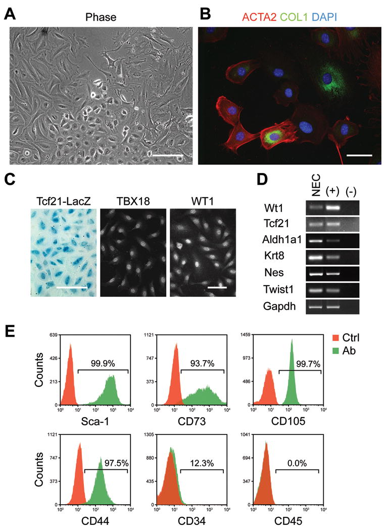
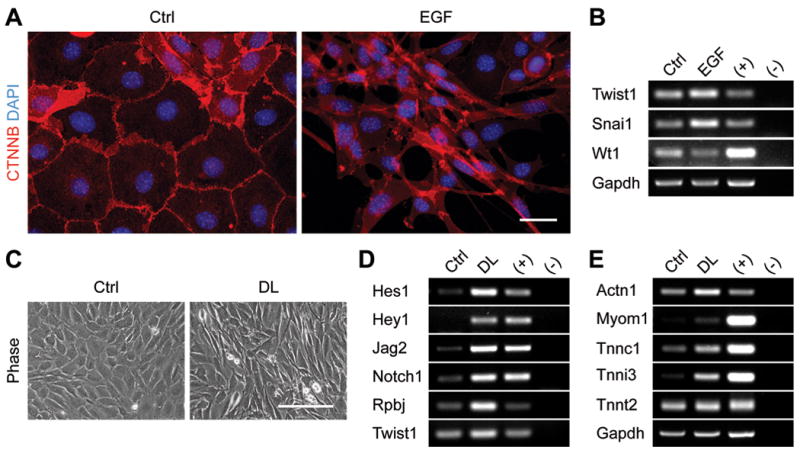
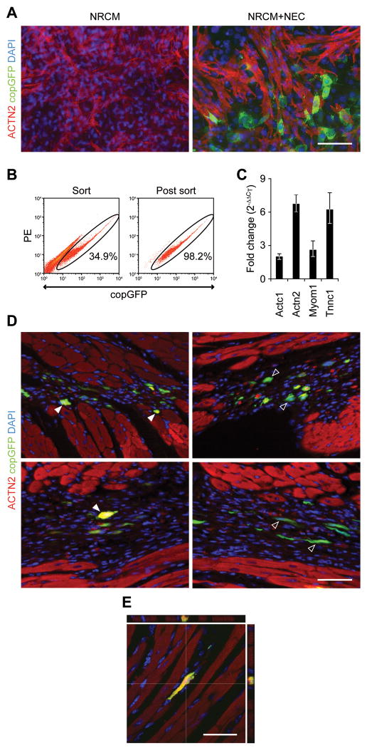
Methods and results: In addition to scattered endothelial and interstitial cells, Notch-activated (EGFP(+)) cells unexpectedly richly populated the adult epicardium. We used fluorescence-activated cell sorting to isolate EGFP(+) cells and excluded hematopoietic (CD45(+)) and endothelial (CD31(+)) subsets. We analyzed EGFP(+)/CD45⁻/CD31⁻ cells, a small (<2%) but distinct subpopulation, by gene expression profiling and functional analyses. We called this mixed cell pool, which had dual multipotent stromal cell and epicardial lineage signatures, Notch-activated epicardial-derived cells (NECs). Myocardial infarction and thoracic aortic banding amplified the NEC pool, increasing fibroblast differentiation. Validating the functional vitality of clonal NEC lines, serum growth factors triggered epithelial-mesenchymal transition and the immobilized Notch ligand Delta-like 1-activated downstream target genes. Moreover, cardiomyocyte coculture and engraftment in NOD-SCID (nonobese diabetic-severe combined immunodeficiency) mouse myocardium increased cardiac gene expression in NECs.

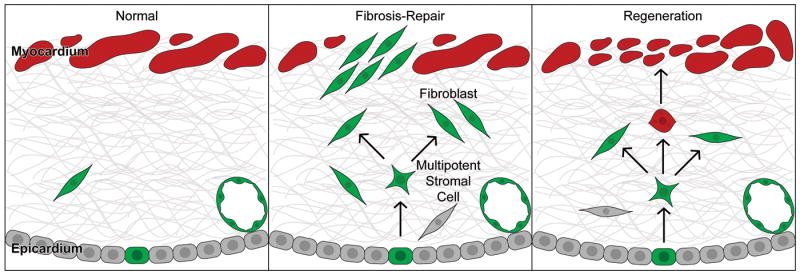
Conclusions: A dynamic Notch injury response activates adult epicardium, producing a multipotent cell population that contributes to fibrosis repair.


Figures

Figure 1
Figure 1. A dynamic and pro-fibrotic epicardial injury response marked by Notch activity
A, EGFP IHC (green) of TNR hearts (apex, left and right ventricles) of control, LAD-MI and TAB (day 7), counterstained for Troponin-I (red) and DAPI (blue) (bar=40 μm) (BZ=borderzone). B, Percentage of EGFP+/Cd31-/Cd45- cells by FACS profiling, 7 days after LAD-MI or TAB. C, Time course of increased EGFP+/CD45-/CD31- cells following LAD-MI and TAB. D, Comparison of gene numbers and categories coordinately up- or downregulated in NECs from LAD-MI and TAB hearts (day 7). E, Independent Q-PCR confirming microarray results.
Figure 2
Figure 2. Adult Notch-activated epicardial-derived cells are multipotent stromal cells
A, Comparison of genes commonly expressed in NECs, adult and embryonic cardiac fibroblasts (ACF and ECF). B, FACS profile of Hoechst stained TNR heart cells to distinguish side population cells and NECs. C, Three-way comparison of of genes commonly expressed in NECs, bone marrow-derived multipotent stromal cells (MSCs) and ACFs. D, Expression of cell surface markers by FACS profiling in NECs (% of cell population positive for marker). E, Co-localization of EGFP and Tcf21-LacZ IHC to annulus fibrosis of adult double transgenic mouse heart (bar = 40 μm).
Figure 3
Figure 3. Confirmation of epicardial and multipotent phenotype in Adult Notch-activated epicardial-derived cell clones
A, Heterogeneity of cell types in NEC clones demonstrated by phase contrast microscopy (bar = 200 μm). B, Cell type heterogeneity in NEC clones demonstrated by ICC for smooth muscle actin (red) and collagen I (green) with DAPI (bar = 40 μm). C, Expression of epicardial markers in NEC clone from TNR/Capsulin-LacZ double reporter mouse by LacZ histochemistry (bar = 200 μm) and TBX18 and WT1 immunostaining (bar = 40 μm). D, Expression of epicardial mRNAs in a NEC clone by RT-PCR. E, Expression of cell surface markers by FACS profiling in NEC clone.
Figure 4
Figure 4. Adult Notch-activated epicardial-derived cell clones undergo epithelial-mesenchymal transition and display intact Notch signaling cascade
A, Epithelial-to-mesenchymal morphological change and β-catenin (red) relocalization induced by EGF in NEC clone, with nuclei stained by DAPI (bar = 40 μm). B, Reciprocal gene regulation of Twist1 and Snai1 (mesenchymal markers) versus Wt1 (epithelial marker) in NECs treated with EGF. C, Morphological changes induced by immobilized Delta-like 1 (DL) in NEC clones demonstrated by phase contrast microscopy (bar = 200 μm). D, Activation of Notch target genes (Hes1, Hey1, Jag2, Notch1, Rbpj), EMT marker (Twist1), and (E) cardiac genes (Actn1, Myom1, and troponins) by immobilized Delta-like ligand in an NEC clone by RT-PCR.
Figure 5
Figure 5. Adult Notch-activated epicardial-derived cells express cardiac genes after NRCM co-culture or engraftment in ventricular myocardium
A, Double immunostaining for copGFP (green) and α-actinin (red), with DAPI staining of nuclei (blue), of two month-old primary newborn rat heart/NRCM cultures, alone or co-cultured with copGFP-NECs (bar = 40 μm). B, Scattergram of copGFP-NECs retrieved from long-term co-cultures by FACS and the post sort confirming purity. C, Q-PCR using mouse-specific primers demonstrating increased expression of cardiac genes in copGFP-NECs retrieved from NRCM co-cultures compare to control cells. D, Identification of copGFP-NECs colonies engrafted in NOD-SCID ventricular myocardium (day 7 post-injection) by double staining for α-actinin (red) and copGFP (green) (bar = 40 μm). E, Expression of α-actinin by engrafted copGFP-NEC as in (D) by confocal microscopy (bar = 40 μm).
Figure 6
Figure 6. A dynamic adult epicardial injury response marked by Notch activity contributes to fibrosis repair
Cartoon model of Notch-activated (CBF1REx4-EGFP+) epicardial-mesothelial cell that delaminates into the sub-epicardial space and undergoes EMT to produce a multipotent stromal cell (MSC) capable of differentiating into fibroblasts (FB), smooth muscle cells (SMC) and other cells, perhaps even cardiomyocytes.

Comment in

  • Kicking the epicardium up a notch.
    Rentschler S, Epstein JA. Rentschler S, et al. Circ Res. 2011 Jan 7;108(1):6-8. doi: 10.1161/CIRCRESAHA.110.237297. Circ Res. 2011. PMID: 21212389 Free PMC article. No abstract available.

Similar articles

Cited by

References

    1. Krenning G, Zeisberg EM, Kalluri R. The origin of fibroblasts and mechanism of cardiac fibrosis. J Cell Physiol - PMC - PubMed
    1. Zeisberg EM, Tarnavski O, Zeisberg M, Dorfman AL, McMullen JR, Gustafsson E, Chandraker A, Yuan X, Pu WT, Roberts AB, Neilson EG, Sayegh MH, Izumo S, Kalluri R. Endothelial-to-mesenchymal transition contributes to cardiac fibrosis. Nat Med. 2007;13:952–961. - PubMed
    1. Snider P, Standley KN, Wang J, Azhar M, Doetschman T, Conway SJ. Origin of cardiac fibroblasts and the role of periostin. Circ Res. 2009;105:934–947. - PMC - PubMed
    1. Zhou B, von Gise A, Ma Q, Hu YW, Pu WT. Genetic fate mapping demonstrates contribution of epicardium-derived cells to the annulus fibrosis of the mammalian heart. Dev Biol. 338:251–261. - PMC - PubMed
    1. Popescu LM, Gherghiceanu M, Manole CG, Faussone-Pellegrini MS. Cardiac renewing: Interstitial cajal-like cells nurse cardiomyocyte progenitors in epicardial stem cell niches. J Cell Mol Med. 2009;13:866–886. - PMC - PubMed

Publication types

MeSH terms

Substances