. 2011 Feb;31(2):360-7.
doi: 10.1161/ATVBAHA.110.212993. Epub 2010 Nov 24.

Formin homology domain-containing protein 1 regulates smooth muscle cell phenotype

Affiliations

Formin homology domain-containing protein 1 regulates smooth muscle cell phenotype

Dean P Staus et al. Arterioscler Thromb Vasc Biol. 2011 Feb.

Abstract

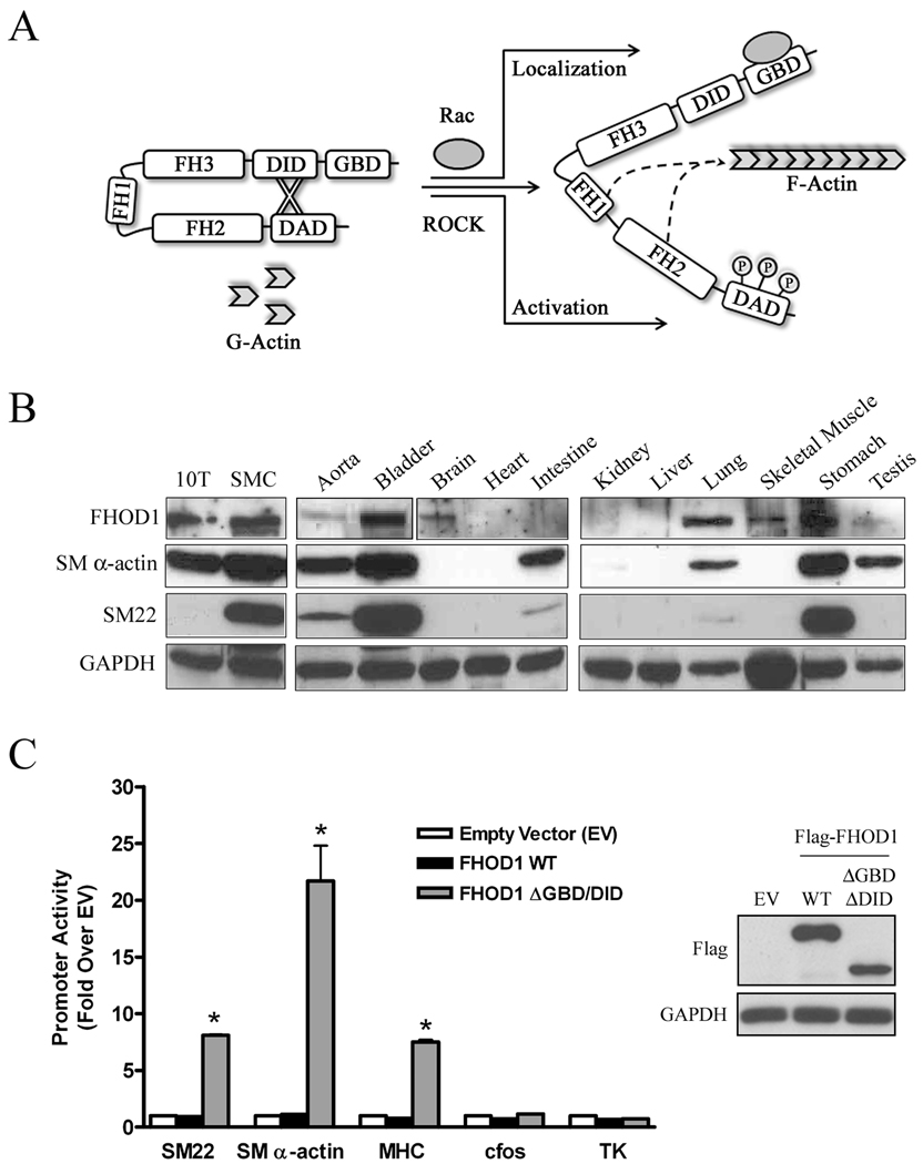
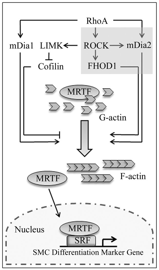
Objective: Our goal was to test whether formin homology protein 1 (FHOD1) plays a significant role in the regulation of smooth muscle cell (SMC) differentiation and, if so, whether Rho kinase (ROCK)-dependent phosphorylation in the diaphanous autoinhibitory domain is an important signaling mechanism that controls FHOD1 activity in SMC.

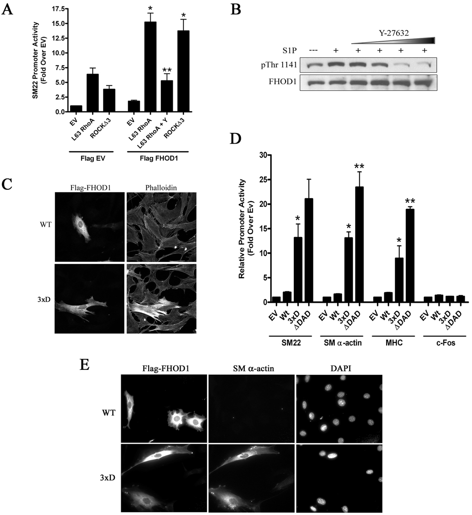
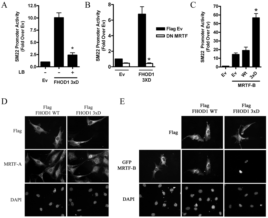
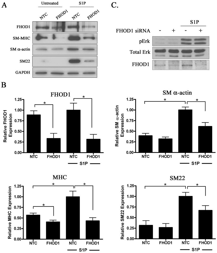
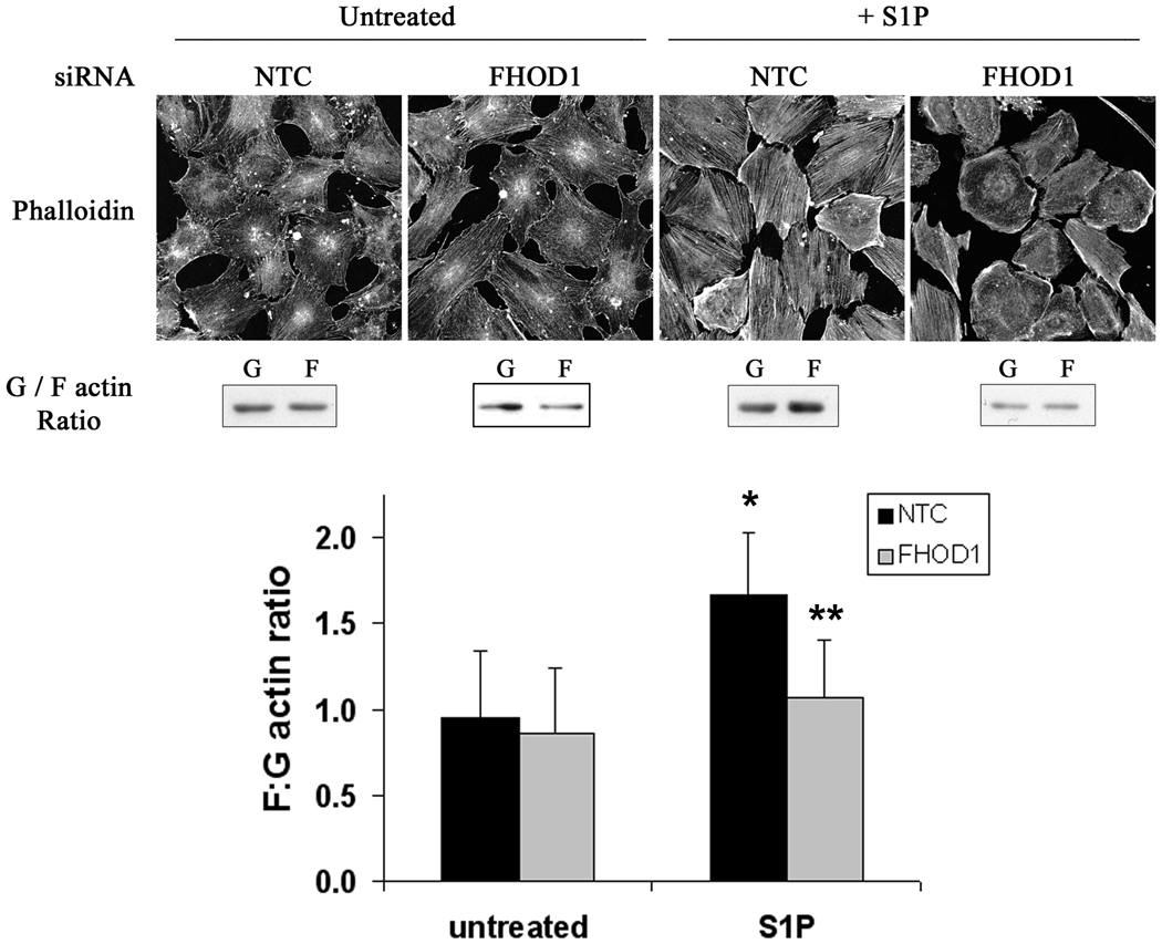
Methods and results: FHOD1 is highly expressed in aortic SMCs and in tissues with a significant SMC component. Exogenous expression of constitutively active FHOD1, but not wild-type, strongly activated SMC-specific gene expression in 10T1/2 cells. Treatment of SMC with the RhoA activator sphingosine-1-phosphate increased FHOD1 phosphorylation at Thr1141, and this effect was completely prevented by inhibition of ROCK with Y-27632. Phosphomimetic mutations to ROCK target residues enhanced FHOD1 activity, suggesting that phosphorylation interferes with FHOD1 autoinhibition. Importantly, knockdown of FHOD1 in SMC strongly inhibited sphingosine-1-phosphate-dependent increases in SMC differentiation marker gene expression and actin polymerization, suggesting that FHOD1 plays a major role in RhoA-dependent signaling in SMC.

Conclusions: Our results indicate that FHOD1 is a critical regulator of SMC phenotype and is regulated by ROCK-dependent phosphorylation. Thus, additional studies on the role of FHOD1 during development and the progression of cardiovascular disease will be important.


Figures

Figure 1
Figure 1. FHOD1 is expressed in SMC-containing tissues and stimulated SMC-specific gene transcription
A) Molecular regulation of FHOD1. ROCK-dependent phosphorylation of three conserved residues within the DAD domain activates FHOD1 while binding to Rac regulates FHOD1 subcellular localization. B) Western Blot of FHOD1, SM α-actin, SM22, and GAPDH expression in the indicated cell-lines and tissues from adult C57/Bl6 mice. C) Flag-tagged wild-type (WT) or ΔGBD/DID FHOD1 variants were transfected into 10T1/2 cells along with luciferase reporter constructs driven by the indicated promoter. Luciferase activity or was analyzed at 24h and is expressed relative to plus empty expression vector (EV). Protein expression (inset), * p<0.05 versus plus empty vector.
Figure 2
Figure 2. ROCK-dependent phosphorylation of FHOD1 activated SMC-specific gene expression
A) SM22 luciferase and constitutively active RhoA (L63) or ROCK (Δ3) were transfected into 10T1/2 cells plus/minus flag-FHOD1. In some experiments cells were treated with Y-27632 (Y) for 8h prior to luciferase determination. * p<0.05 versus minus FHOD1, ** p<0.05 versus L63 RhoA + FHOD1. B) Primary mouse SMCs were serum starved for 24h and then pre-treated with increasing amounts of Y-27632 (10, 25, 50, 100μM) for 90 m and the phosphatase inhibitor, calyculin A (50nM), for 5 m. After addition of S1P (10μM), FHOD1 phosphorylation was measured at 7.5 m using a phospho-specific antibody against Thr-1141. C and E) 10T1/2 cells were transfected with WT FHOD1 or an FHOD1 variant containing phosphomimetic mutations to the three ROCK target residues within the auto-inhibitory domain (3xD). Cells were serum starved in 0.5% FBS containing media for 24h, and then stained for actin polymerization (C) or endogenous SM α-actin expression (E). D) The indicated flag-FHOD1 variants and luciferase reporters were transfected into 10T1/2 cells and luciferase activity was measured at 24h. * p<0.05 versus Wt. ** p<0.05 versus 3xD.
Figure 3
Figure 3. FHOD1 regulated MRTF sub-cellular localization and activity
A) 10T1/2 cells were transfected with the 3xD FHOD1 variant and SM22 luciferase. Some cells were treated with 10μM latrunculin B (LB) for 6h prior to luciferase determination. * p<0.05 versus untreated. B) The 3xD FHOD1 variant was co-transfected with dominant negative MRTF and SM22 luciferase. * p<0.05 versus FHOD1 plus EV. C) SM22 luciferase, MRTF-B, and either WT or 3xD FHOD1 were co-transfected into 10T1/2 cells. * p<0.05 versus Wt FHOD1 plus MRTF-B. D and E) Localization of endogenous MRTF-A (D) or GFP-MRTF-B (E) in serum starved 10T1/2 cells co-expressing Wt or 3xD FHOD1.
Figure 4
Figure 4. Knock-down of FHOD1 in SMC inhibited SMC differentiation marker gene expression
A) Primary aortic SMC were transfected with non-targeted control (NTC) or FHOD1 siRNAs for 72h. Following 24h of complete serum starvation, cells were treated with S1P for an additional 24h. Lysates were subjected to Western Blot analysis using the indicated antibody. B) Quantification of FHOD1 and SM marker gene expression from five independent experiments. Protein expression was normalized to GAPDH expression and is expressed relative to NTC cells treated with S1P set to 1. * p<0.05. C) Control and FHOD1 knockdown SMC were treated with S1P for 12.5 minutes. ERK activity was then measured by Western Blotting using a phospho-specific ERK antibody.
Figure 5
Figure 5. Knock-down of FHOD1 inhibited S1P-dependent actin polymerization
SMCs were plated on 10μg/mL fibronectin, depleted of FHOD1 by siRNA for 72h, serum starved for 24h, and treated with S1P for 30 min. Actin polymerization was measured by phalloidin staining (top panels) and actin sedimentation assays (middle panels; see methods for more details). The results of three separate actin sedimentation experiments are quantified in the bottom panel. * p<0.05 versus untreated NTC; ** p<0.05 versus S1P-treated NTC.
Figure 6
Figure 6. The RhoA-dependent signaling pathways that regulate SMC differentiation
RhoA activates mDia1, mDia2, and ROCK through direct binding. ROCK activity stimulates actin polymerization through LIM-kinase-mediated inhibition of cofilin. Our recent results (shaded box) suggest that ROCK also enhances the activities of FHOD1 and mDia2 through direct phosphorylation of the DAD auto-regulatory domains of these proteins. Collectively these pathways reduce cellular G-actin pools leading to MRTF nuclear accumulation, and our data suggest that all three DRFs are required for full RhoA-dependent activation of SMC-specific gene expression.

Similar articles

Cited by

References

    1. Owens GK, Kumar MS, Wamhoff BR. Molecular regulation of vascular smooth muscle cell differentiation in development and disease. Physiol Rev. 2004;84:767–801. - PubMed
    1. Landerholm TE, Dong XR, Lu J, Belaguli NS, Schwartz RJ, Majesky MW. A role for serum response factor in coronary smooth muscle differentiation from proepicardial cells. Development. 1999;126:2053–2062. - PubMed
    1. Niu Z, Yu W, Zhang SX, Barron M, Belaguli NS, Schneider MD, Parmacek M, Nordheim A, Schwartz RJ. Conditional mutagenesis of the murine serum response factor gene blocks cardiogenesis and the transcription of downstream gene targets. J Biol Chem. 2005;280:32531–32538. - PubMed
    1. Li S, Chang S, Qi X, Richardson JA, Olson EN. Requirement of a myocardin-related transcription factor for development of mammary myoepithelial cells. Mol Cell Biol. 2006;26:5797–5808. - PMC - PubMed
    1. Li J, Zhu X, Chen M, Cheng L, Zhou D, Lu MM, Du K, Epstein JA, Parmacek MS. Myocardin-related transcription factor B is required in cardiac neural crest for smooth muscle differentiation and cardiovascular development. Proc Natl Acad Sci U S A. 2005;102:8916–8921. - PMC - PubMed

Publication types

MeSH terms