. 2011 Feb 3;117(5):1700-6.
doi: 10.1182/blood-2010-08-300301. Epub 2010 Nov 24.

Fibrinopeptides A and B release in the process of surface fibrin formation

Affiliations

Fibrinopeptides A and B release in the process of surface fibrin formation

Tomas Riedel et al. Blood. .

Abstract

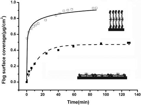
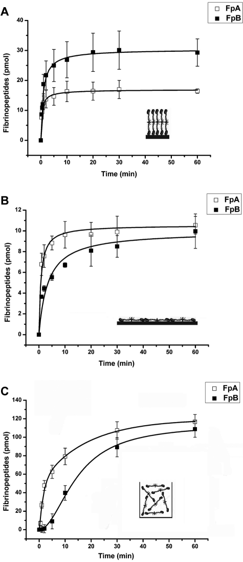
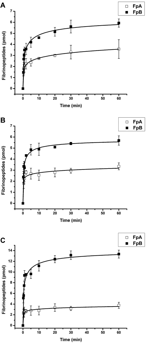
Fibrinogen adsorption on a surface results in the modification of its functional characteristics. Our previous studies revealed that fibrinogen adsorbs onto surfaces essentially in 2 different orientations depending on its concentration in the solution: "side-on" at low concentrations and "end-on" at high concentrations. In the present study, we analyzed the thrombin-mediated release of fibrinopeptides A and B (FpA and FpB) from fibrinogen adsorbed in these orientations, as well as from surface-bound fibrinogen-fibrin complexes prepared by converting fibrinogen adsorbed in either orientation into fibrin and subsequently adding fibrinogen. The release of fibrinopeptides from surface-adsorbed fibrinogen and from surface-bound fibrinogen-fibrin complexes differed significantly compared with that from fibrinogen in solution. The release of FpB occurred without the delay (lag phase) characteristic of its release from fibrinogen in solution. The amount of FpB released from end-on adsorbed fibrinogen and from adsorbed fibrinogen-fibrin complexes was much higher than that of FpA. FpB is known as a potent chemoattractant, so its preferential release suggests a physiological purpose in the attraction of cells to the site of injury. The N-terminal portions of fibrin β chains including residues Bβ15-42, which are exposed after cleavage of FpB, have been implicated in many processes, including angiogenesis and inflammation.


Figures

Figure 1
Figure 1
Adsorption of a primary fibrinogen layer observed by ATR FT-IR spectroscopy. Fibrinogen (Fbg) was adsorbed onto polystyrene from high (500 μg/mL)– and low (20 μg/mL)–concentration fibrinogen solutions (□ and ■, respectively). Inserts show an illustrative arrangement of fibrinogen molecules adsorbed in the end-on (top insert) and side-on orientations.
Figure 2
Figure 2
SPR-observed adsorption of primary and secondary fibrinogen layers. A primary layer was formed by the adsorption of fibrinogen onto the gold surface, followed by washing with TBS and PBS; a secondary fibrinogen layer was formed by the immobilization of fibrinogen onto the primary layer, which was treated with thrombin to convert the adsorbed fibrinogen into fibrin. Arrows indicate the replacement of solutions. Curve 1, fibrinogen (Fbg) 500 μg/mL, TBS, PBS; thrombin 2.5 U/mL (THR), PBS, TBS; mixture of PPACK 10μM and hirudin 6 U/mL, TBS; fibrinogen 200 μg/mL, TBS. Curve 2, fibrinogen 20 μg/mL, TBS, PBS; thrombin 2.5 U/mL, PBS, TBS; PPACK 10μM, hirudin 6 U/mL, TBS; fibrinogen 200 μg/mL, TBS. Curve 3, fibrinogen 20 μg/mL, TBS, PBS, TBS; fibrinogen 200 μg/mL, TBS. Curve 4, fibrinogen 20 μg/mL, TBS, PBS; thrombin 2.5 U/mL, PBS, TBS; PPACK 10 μM and hirudin 6 U/mL, TBS; fibrinogen 20 μg/mL, TBS.
Figure 3
Figure 3
Morphology of fibrinogen-fibrin layers on carbon as observed by TEM. Fibrinogen was adsorbed on carbon-coated mica at a concentration of 20 μg/mL for 150 minutes (A) and 500μg/mL for 30 minutes (B), treated with thrombin at 2.5 U/mL for 60 minutes, a mixture of PPACK at 10μM and hirudin at 6 U/mL for 20 minutes, and fibrinogen at a concentration of 200 μg/mL for 60 minutes. The layers were contrasted with uranyl acetate, dehydrated with a series of water-ethanol solutions, and dried.
Figure 4
Figure 4
Kinetics of thrombin-mediated release of fibrinopeptides from the primary fibrinogen layers adsorbed on polystyrene. The amounts of FpA (□) and FpB (■) released from the primary fibrinogen layers adsorbed from the high (500 μg/mL, 30 minutes) and low (20 μg/mL, 150 minutes) fibrinogen concentrations (A and B, respectively) and from fibrinogen in solution (C) was determined by HPLC, as described in “Methods.” The concentration of thrombin in all experiments was 2.5 U/mL. Each point represents a mean value obtained from 3 independent experiments in which the total amount released in one well at the indicated time interval was measured. Inserts in panels a and b show an illustrative arrangement of fibrinogen molecules adsorbed in the end-on and side-on orientations, respectively, and in panel C fibrinogen in solution.
Figure 5
Figure 5
Kinetics of thrombin-mediated release of fibrinopeptides from the secondary layer of surface-bound fibrinogen-fibrin complexes. All fibrinogen-fibrin complexes were formed by the immobilization of the secondary fibrinogen layer on thrombin-treated (2.5 U/mL of thrombin for 60 minutes, followed by a mixture of PPACK and hirudin for 20 minutes) primary fibrinogen layers adsorbed onto polystyrene. (A) The primary layer was adsorbed from 500 μg/mL of fibrinogen for 30 minutes; the secondary layer was immobilized at 200 μg/mL of fibrinogen for 60 minutes. (B) The primary layer was adsorbed from 20 μg/mL of fibrinogen for 150 minutes; the secondary layer was immobilized at 20 μg/mL of fibrinogen for 120 minutes. (C) The primary layer was adsorbed from 20 μg/mL of fibrinogen for 150 minutes; the secondary layer was immobilized at 200 μg/mL of fibrinogen for 60 minutes. The amounts of FpA (□) and FpB (■) released from the secondary layers upon incubation with 2.5 U/mL of thrombin were determined by HPLC, as described in “Methods.” Each point represents a mean value obtained from 3 independent experiments.

Similar articles

Cited by

References

    1. Geer CB, Tripathy A, Schoenfisch MH, Lord ST, Gorkun OV. Role of B-b knob-hole interactions in fibrin binding to adsorbed fibrinogen. J Thromb Haemost. 2007;5(12):2344–2351. - PubMed
    1. Geer CB, Rus IA, Lord ST, Schoenfisch MH. Surface-dependent fibrinopeptide A accessibility to thrombin. Acta Biomater. 2007;3(5):663–668. - PubMed
    1. Evans-Nguyen KM, Fuierer RR, Fitchett BD, Tolles LR, Conboy JC, Schoenfisch MH. Changes in adsorbed fibrinogen upon conversion to fibrin. Langmuir. 2006;22(11):5115–5121. - PubMed
    1. Soman P, Rice Z, Siedliecki CA. Measuring the time-dependent functional activity of adsorbed fibrinogen by atomic force microscopy. Langmuir. 2008;24(16):8801–8806. - PubMed
    1. Savage B, Ruggeri ZM. Selective recognition of adhesive sites in surface-bound fibrinogen by glycoprotein IIb-IIIa on nonactivated platelets. J Biol Chem. 1991;266(17):11227–11233. - PubMed

Publication types