. 2010 Dec;30(4):328-39.
doi: 10.1097/WNO.0b013e3181f7f833.

Oral resveratrol reduces neuronal damage in a model of multiple sclerosis

Affiliations

Oral resveratrol reduces neuronal damage in a model of multiple sclerosis

Kenneth S Shindler et al. J Neuroophthalmol. 2010 Dec.

Abstract

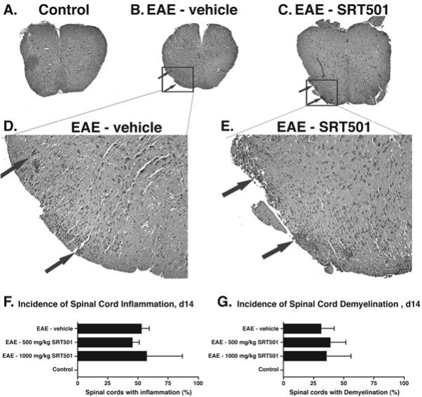
Background: Neuronal loss in multiple sclerosis (MS) and its animal model, experimental autoimmune encephalomyelitis (EAE), correlates with permanent neurological dysfunction. Current MS therapies have limited the ability to prevent neuronal damage.

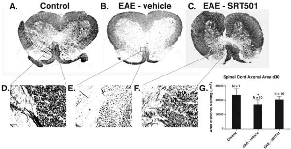
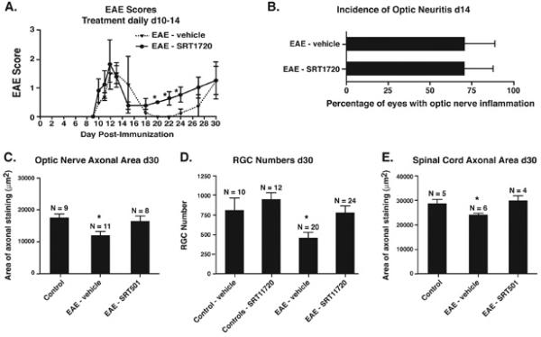
Methods: We examined whether oral therapy with SRT501, a pharmaceutical grade formulation of resveratrol, reduces neuronal loss during relapsing-remitting EAE. Resveratrol activates SIRT1, an NAD+-dependent deacetylase that promotes mitochondrial function.

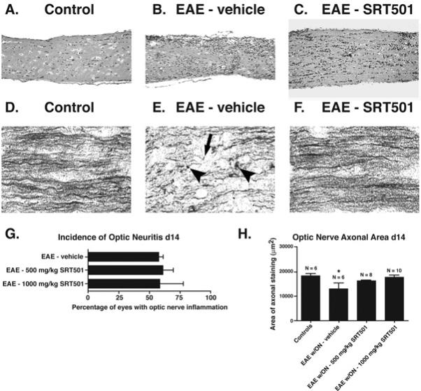
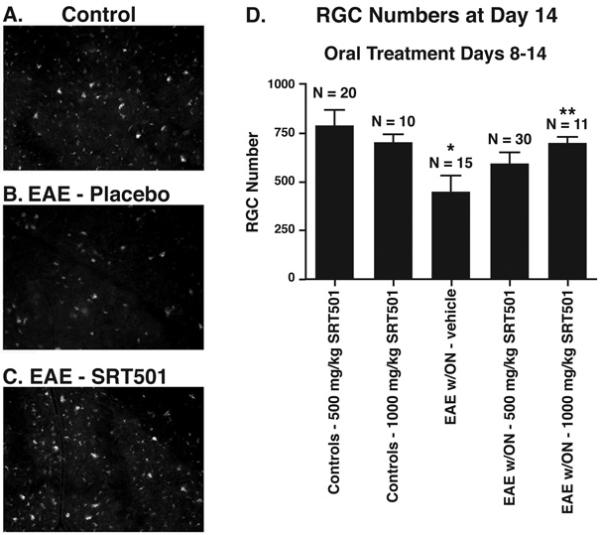
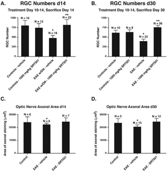
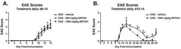
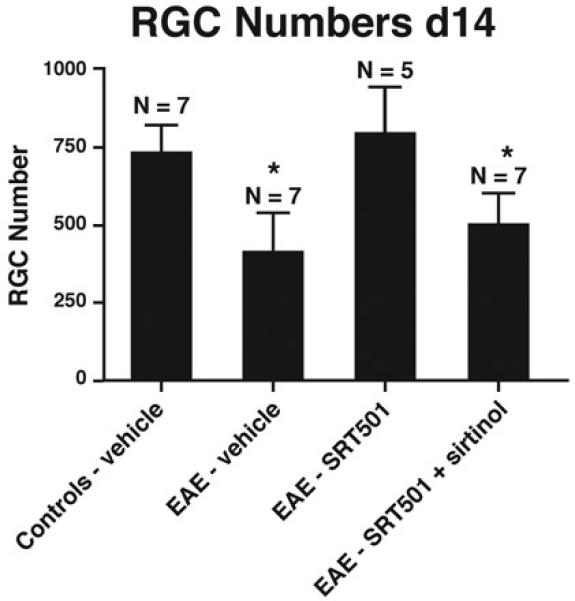
Results: Oral SRT501 prevented neuronal loss during optic neuritis, an inflammatory optic nerve lesion in MS and EAE. SRT501 also suppressed neurological dysfunction during EAE remission, and spinal cords from SRT501-treated mice had significantly higher axonal density than vehicle-treated mice. Similar neuroprotection was mediated by SRT1720, another SIRT1-activating compound; and sirtinol, an SIRT1 inhibitor, attenuated SRT501 neuroprotective effects. SIRT1 activators did not prevent inflammation.

Conclusions: These studies demonstrate that SRT501 attenuates neuronal damage and neurological dysfunction in EAE by a mechanism involving SIRT1 activation. SIRT1 activators are a potential oral therapy in MS.


Figures

Fig. 1
Fig. 1
Oral SRT501 reduces axonal damage during optic neuritis without suppressing inflammation. A. A longitudinal section from a control optic nerve stained by H&E shows normal histology. B. A representative optic nerve from an EAE mouse 14 days post-immunization stained by H&E demonstrates increased cellularity from infiltrating inflammatory cells observed during optic neuritis. C. H&E staining of a day 14 optic nerve from an EAE mouse treated with 1000 mg/kg SRT501 daily from d8–14 shows inflammatory infiltrates are still present. D. Normal axonal staining by Bielschowsky silver impregnation in a control optic nerve. E. Axonal damage marked by terminal axonal ovoids (arrowheads) and truncated axons (arrow) found in a nerve with optic neuritis 14 days after induction of EAE. F. Normal optic nerve axonal staining in an eye with optic neuritis from an EAE mouse treated with 1000 mg/kg SRT501 d8–14. G. The percentage of eyes that developed optic neuritis in EAE mice treated with 500 or 1000 mg/kg SRT501 daily from d8–14 was similar to the incidence of optic neuritis in EAE mice treated with vehicle (mean ± SEM of 3 experiments). H. Axonal density, measured as the area of positive silver staining (mean ± SEM), was significantly decreased in eyes with optic neuritis compared to either control eyes or optic neuritis eyes from EAE mice treated with 1000 mg/kg SRT501 daily from d8–14 (*p < 0.05). One experiment of three is shown. Original magnification 20× (AC) and 40× (D–F).
Fig. 2
Fig. 2
Oral SRT501 treatment from d8–14 post-immunization prevents RGC loss in EAE optic neuritis eyes. A. Numerous fluorescently-labeled RGCs from a representative field in a control retina. B. Fewer RGCs are seen in a corresponding area of retina in an EAE eye with optic neuritis. C. Retina from an optic neuritis eye in an EAE mouse treated with 1000 mg/kg SRT501 contains numerous RGCs similar to controls. D. Optic neuritis induced significant loss of RGCs compared to control eyes (*p < 0.05). 500 mg/kg SRT501 lead to a non-significant trend of increased RGC survival, and 1000 mg/kg SRT501 lead to significantly higher numbers of surviving RGCs in optic neuritis eyes compared to vehicle (**p < 0.05). N = number of eyes. Data represent the mean ± SEM number of RGCs counted in 12 standardized fields/eye.
Fig. 3
Fig. 3
Oral SRT501 treatment beginning after onset of optic neuritis, from d10–14 post-immunization, prevents acute axon and RGC loss at day 14 and maintains these neuroprotective effects at day 30. A. The significant loss of RGCs detected in EAE optic neuritis eyes (*p < 0.01 vs. controls) at day 14 is prevented by 1000 mg/kg SRT501 (**p < 0.01 vs. vehicle-treated EAE optic neuritis eyes). B. Similar RGC loss is detected in eyes from vehicle-treated EAE mice at day 30 (*p < 0.01 vs. controls), with increased RGC survival in SRT501-treated EAE eyes (**p < 0.001 vs. vehicle-treated EAE eyes). C. Decreased area of axonal silver staining in EAE optic neuritis eyes compared to controls at day 14 (*p < 0.05) was prevented by 1000 mg/kg SRT501. D. 1000 mg/kg SRT501 also attenuated axonal loss detected in eyes from EAE mice at day 30 (*p < 0.01). One representative experiment of three is shown.
Fig. 4
Fig. 4
SRT501 suppresses EAE remission but not acute exacerbations. A. No difference was found between the degree of EAE in mice treated with vehicle (n = 19), 500 mg/kg SRT501 (n = 30), or 1000 mg/kg SRT501 (n = 13) given by oral gavage daily from d8–14 up to the peak of disease at sacrifice on day 14. B. In an extended treatment study, no difference in development of acute EAE was detected between vehicle-treated (n = 5) and 1000 mg/kg SRT501-treated (n = 6) EAE mice treated d10–14. During remission at days 20–25, EAE was significantly suppressed in SRT501-treated vs. vehicle-treated mice (*p < 0.05). One of three experiments is shown.
Fig. 5
Fig. 5
SRT501 does not reduce spinal cord inflammation or demyelination at day 14 post-immunization. A. An H&E stained cross-section through the spinal cord of a control mouse demonstrates normal histology at 4× original magnification. B. Focal areas containing inflammatory cell infiltrates (arrows) are shown in a spinal cord from an EAE mouse. C. Similar foci of inflammation (arrows) are present in the spinal cord of an EAE mouse treated with 1000 mg/kg SRT501 daily from d8–14. D. Higher magnification (40×) photograph of areas of inflammation shown in B. E. Higher magnification (40×) photograph of areas of inflammation shown in C. F. The percentage of spinal cord sections that contained inflammation in EAE mice treated with 500 or 1000 mg/kg SRT501 daily from d8–14 was similar to the incidence in EAE mice treated with vehicle (mean ± SEM of 3 experiments), with no inflamed spinal cords found in control mice. G. The percentage of spinal cord sections that contained demyelination, assessed by LFB staining, in EAE mice treated with 500 or 1000 mg/kg SRT501 daily from d8–14 was similar to the incidence in EAE mice treated with vehicle (mean ± SEM of 3 experiments), with no demyelination found in control mice.
Fig. 6
Fig. 6
SRT501 treatment during acute inflammation on d10–14 prevents loss of axons in EAE spinal cord 30 days post-immunization. A. Bielschowsky silver stained section of control spinal cord shows normal axonal staining. Higher magnification of boxed area is shown in D, with normal gray matter (left half) and medial corticospinal tract (right half) demonstrating high density of silver stained axons. B. Spinal cord from an EAE mouse contains focal areas of axonal loss (arrowheads) and mild diffuse decrease in silver staining. Higher magnification of boxed area shown in E demonstrates a focal area of medial corticospinal tract with notable decrease in axons compared to control spinal cord. C. Normal axonal staining observed in the spinal cord of an EAE mouse treated with 1000 mg/kg SRT501. Higher magnification of boxed area shown in F demonstrates higher axonal concentration than vehicle-treated mouse spinal cord. G. Quantification of axonal staining demonstrates that significant axonal loss compared to controls occurs in spinal cords from vehicle-treated (*p < 0.001), but not SRT501-treated, EAE mice. One of three experiments is shown. Photographs A–C shown at 4× original magnification, D–F at 40× original magnification.
Fig. 7
Fig. 7
SRT1720 treatment exhibits similar neuroprotective effects in EAE as SRT501, without reduction of inflammation. A. No difference in development of acute EAE was detected between vehicle-treated (n = 5) and 100 mg/kg SRT1720-treated (n = 5) EAE mice treated d10–14. During remission at days 20–24, EAE was significantly suppressed in SRT1720-treated vs. vehicle-treated mice (*p < 0.05). One of two experiments is shown. B. The incidence of optic neuritis (mean ± SEM of 3 experiments) did not differ between vehicle-treated and SRT1720-treated EAE mice 14 days post-immunization. C. SRT1720 prevents the significant loss of axons that occurs in EAE optic nerves at day 30 (*p < 0.05). Data represent the mean ± SEM area of positive silver staining measured in one of two experiments. D. SRT1720 also prevents the significant loss of RGCs that occurs in EAE eyes at day 30 (*p < 0.01). Data represent the mean ± SEM number of RGCs measured in each eye. E. SRT1720 has similar neuroprotective effects for spinal cord axons, preventing the significant loss that occurs in EAE spinal cords at day 30 (*p < 0.05). Data represent the mean ± SEM area of positive silver staining measured in one of two experiments.
Fig. 8
Fig. 8
The SIRT1 inhibitor sirtinol reduces neuroprotective effects of SRT501 on RGCs during optic neuritis. 1000 mg/kg SRT501 administered daily from d10–14 in EAE mice results in significantly higher RGC numbers compared to optic neuritis eyes from vehicle-treated EAE mice, but intravitreal injection of sirtinol (100 μM) in SRT501-treated mice on d11 results in decreased RGC numbers (*p < 0.05) following sacrifice on day 14.

Comment in

References

    1. Noseworthy JH, Lucchinetti C, Rodriguez M, Weinshenker BG. Multiple sclerosis. N Eng J Med. 2002;343:938–952. - PubMed
    1. Davie CA, Barker GJ, Webb S, Tofts PS, Thompson AJ, Harding AE, McDonald WI, Miller DH. Persistent functional deficit in multiple sclerosis and autosomal dominant ataxia associated with axon loss. Brain. 1995;118:1583–1592. - PubMed
    1. Losseff NA, Webb SL, O'Riordan JI, Page R, Wang L, Barker GJ, Tofts PS, McDonald WI, Miller DH, Thompson AJ. Spinal cord atrophy and disability in multiple sclerosis: a new reproducible and sensitive MRI method with potential to monitor disease progression. Brain. 1996;119:701–708. - PubMed
    1. Losseff NA, Wang L, Lai HM, Yoo DS, Gawne-Cain ML, McDonald WI, Miller DH, Thompson AJ. Progressive cerebral atrophy in multiple sclerosis: a serial MRI study. Brain. 1996;119:2009–2019. - PubMed
    1. Fisher JB, Jacobs DA, Markowitz CE, Galetta SL, Volpe NJ, Nano-Schiavi ML, Baier ML, Frohman EM, Winslow H, Frohman TC, Calabresi PA, Maguire MG, Cutter GR, Balcer LJ. Relation of visual function to retinal nerve fiber layer thickness in multiple sclerosis. Ophthalmol. 2006;113:324–332. - PubMed

Publication types

MeSH terms