. 2010 Dec;67(6):1745-56; discussion 1756.
doi: 10.1227/NEU.0b013e3181f74105.

An assessment of current brain targets for deep brain stimulation surgery with susceptibility-weighted imaging at 7 tesla

Affiliations

An assessment of current brain targets for deep brain stimulation surgery with susceptibility-weighted imaging at 7 tesla

Aviva Abosch et al. Neurosurgery. 2010 Dec.

Abstract

Background: Deep brain stimulation (DBS) surgery is used for treating movement disorders, including Parkinson disease, essential tremor, and dystonia. Successful DBS surgery is critically dependent on precise placement of DBS electrodes into target structures. Frequently, DBS surgery relies on normalized atlas-derived diagrams that are superimposed on patient brain magnetic resonance imaging (MRI) scans, followed by microelectrode recording and macrostimulation to refine the ultimate electrode position. Microelectrode recording carries a risk of hemorrhage and requires active patient participation during surgery.

Objective: To enhance anatomic imaging for DBS surgery using high-field MRI with the ultimate goal of improving the accuracy of anatomic target selection.

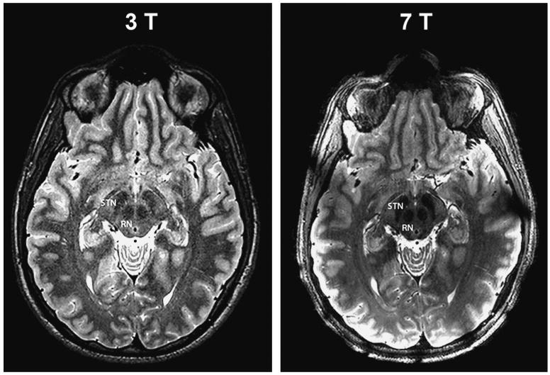
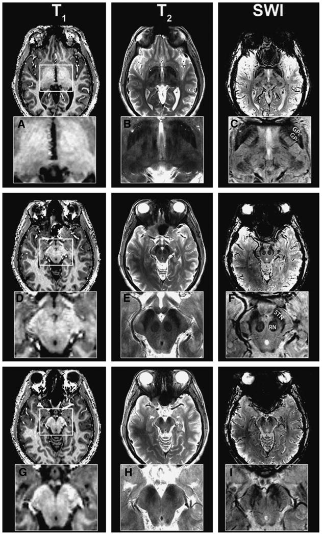
Methods: Using a 7-T MRI scanner combined with an array of acquisition schemes using multiple image contrasts, we obtained high-resolution images of human deep nuclei in healthy subjects.

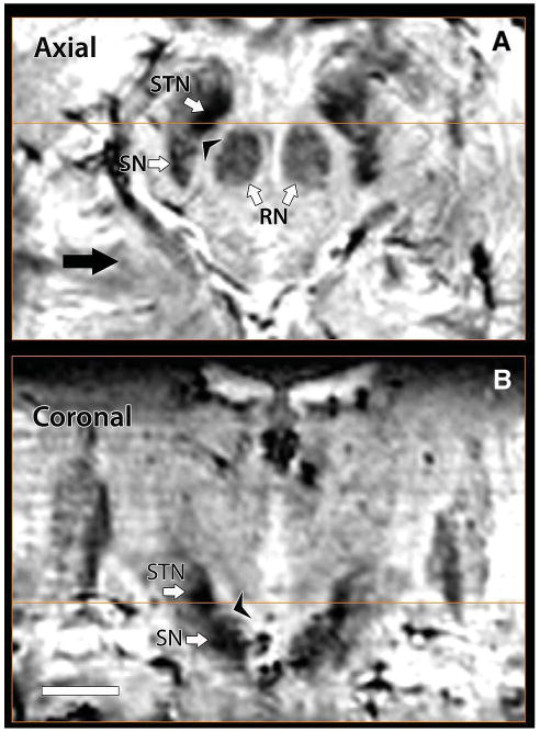
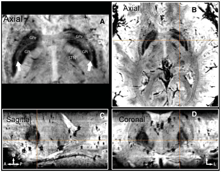
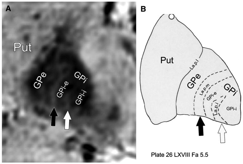
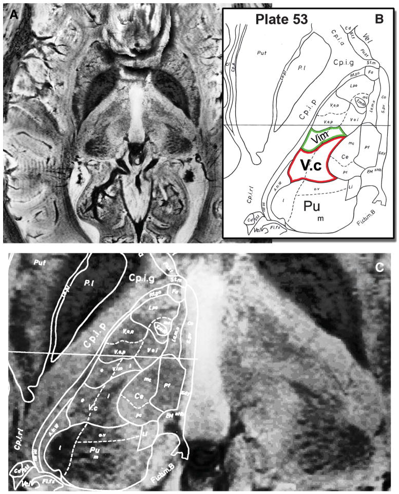
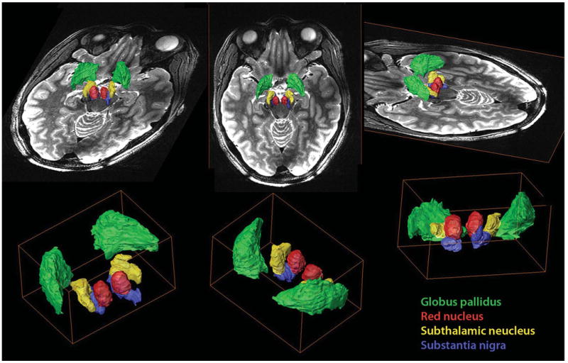
Results: Superior image resolution and contrast obtained at 7 T in vivo using susceptibility-weighted imaging dramatically improved anatomic delineation of DBS targets and allowed the identification of internal architecture within these targets. A patient-specific, 3-dimensional model of each target area was generated on the basis of the acquired images.

Conclusion: Technical developments in MRI at 7 T have yielded improved anatomic resolution of deep brain structures, thereby holding the promise of improving anatomic-based targeting for DBS surgery. Future study is needed to validate this technique in improving the accuracy of targeting in DBS surgery.


Figures

Figure 1
Figure 1
Advantages of higher magnetic field. High-resolution T2-weighted images through the level of the subthalamic nucleus (STN) of a single healthy subject acquired on 3-T and 7-T magnets with matched resolution and acquisition time (0.375 × 0.375 × 2.0 mm3; total acquisition time, 7 min). The 7-T image offers better signal-to-noise ratio (smoother and less grainy) and superior contrast especially at the level of the mid-brain. RN, red nucleus.
Figure 2
Figure 2
Image contrasts at 7 T. Axial brain images obtained with susceptibility-weighted imaging (SWI) at 7 T at the level of globus pallidus interna (GPi) and thalamus (A-C), subthalamic nucleus (STN; D-F), and substantia nigra (SN) caudal to STN (G-I). To emphasize the contrast differences, an inset of the magnifed midbrain region (white box) is shown. T1-weighted images have no obvious identifiable structures (A, D, G); T2-weighted images (B, E, H) delineate major nuclei; SWI images (C, F, I) exhibit an abundance of fine structural details within the same region (see text).
Figure 3
Figure 3
Direct visualization of subthalamic nucleus (STN) and substantia nigra (SN). In 7T susceptibility-weighted imaging axial (A) and coronal (B) images, a boundary between STN and SN can be seen; note also how clearly defined the perimeter of STN appears in both hemispheres. Additionally, examination of red nucleus (RN) reveals a complex internal structure (A).
Figure 4
Figure 4
Direct visualization of globus pallidus interna (GPi) and globus pallidus externa (GPe). Three-plane imaging demonstrating the feasibility of distinguishing GPi from GPe in vivo with susceptibility-weighted imaging at 7 T. GPi is marked and displayed on all 3 orthogonal slices (B-D), immediately caudal to the anterior and posterior commissure line. A, lamina pallidi medialis. The thin layer (arrows) separating GPi from GPe.
Figure 5
Figure 5
In vivo visualization of lamina pallidi incompleta (La.p.i.). Detection of the lamina pallidi incompleta, the border between the internal (GPi-i) and external (GPi-e) segments of globus pallidus interna, is demonstrated. A, a magnified view of the left GP (from Figure 4D). B, the corresponding histologically defined outline (plate 26 from the Schaltenbrand and Wahren atlas). Black arrow points to the border between globus pallidus externa (GPe) and GPi (lamina pallidi medialis [La.p.m.]); white arrow, to the lamina pallidi incompleta. Put, putamen.
Figure 6
Figure 6
Direct visualization of internal thalamic nuclei in vivo. A, an axial susceptibility-weighted imaging slice through the thalamus at the level of the anterior and posterior commissure plane is shown. B, the corresponding histologically defined outline (plate 53 from the Schaltenbrand and Wahren atlas) that is superimposed on the magnetic resonance image in C. Note the clear visualization of the anterior and medial aspects of the pulvinar (Pu), the arrowhead shape of ventral caudalis (Vc; red outline; compare with unmarked right hemisphere), and the image contrast modulation within the thalamus corresponding to Vim (green outline).
Figure 7
Figure 7
A 3-dimensional model of the mesencephalon, thalamus, and surrounding regions. Volume renderings of the globus pallidus (green), red nucleus (red), subthalamic nucleus (yellow), and substantia nigra (blue) fused with a T2-weighted image. Note the clear spatial representation that the model provides regarding the localization, dimensions, and orientation of the key nuclei within the region. The model depicts the main anatomic components in the region and their relationship to the corresponding deep brain stimulation target.

References

    1. Starr PA. Placement of deep brain stimulators into the subthalamic nucleus or globus pallidus internus: technical approach. Stereotact Funct Neurosurg. 2002;79(3-4):118–145. - PubMed
    1. Rijkers K, Temel Y, Visser-Vandewalle V, et al. The microanatomical environment of the subthalamic nucleus: technical note. J Neurosurg. 2007;107(1):198–201. - PubMed
    1. Patel NK, Khan S, Gill SS. Comparison of atlas- and magnetic-resonance-imaging-based stereotactic targeting of the subthalamic nucleus in the surgical treatment of Parkinson's disease. Stereotact Funct Neurosurg. 2008;86(3):153–161. - PubMed
    1. Ashkan K, Blomstedt P, Zrinzo L, et al. Variability of the subthalamic nucleus: the case for direct MRI guided targeting. Br J Neurosurg. 2007;21(2):197–200. - PubMed
    1. Brierley JB, Beck E. The significance in human stereotactic brain surgery of individual variation in the diencephalon and globus pallidus. J Neurol Neurosurg Psychiatry. 1959;22:287–298. - PMC - PubMed

Publication types