

Storable, thermally activated, near-infrared chemiluminescent dyes and dye-stained microparticles for optical imaging
- PMID: 21107365
- PMCID: PMC3043620
- DOI: 10.1038/nchem.871
Storable, thermally activated, near-infrared chemiluminescent dyes and dye-stained microparticles for optical imaging
Abstract
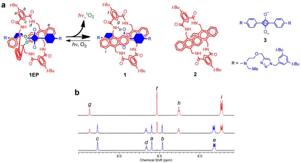
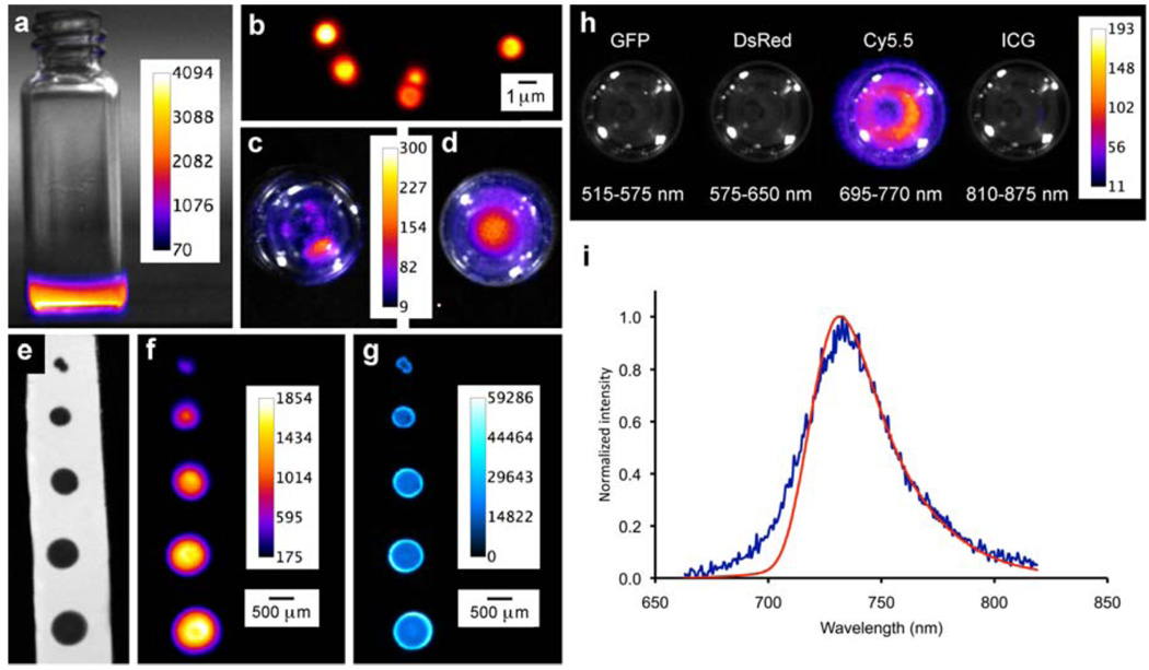
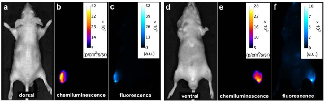
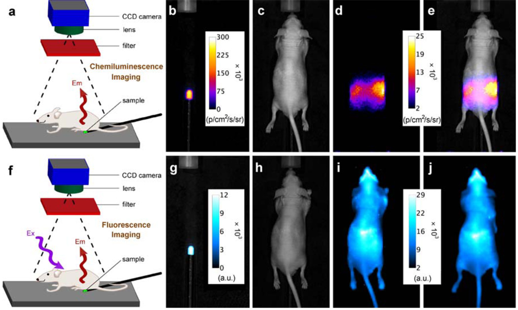
Imaging techniques are a vital part of clinical diagnostics, biomedical research and nanotechnology. Optical molecular imaging makes use of relatively harmless, low-energy light and technically straightforward instrumentation. Self-illuminating, chemiluminescent systems are particularly attractive because they have inherently high signal contrast due to the lack of background emission. Currently, chemiluminescence imaging involves short-lived molecular species that are not stored but are instead generated in situ, and they typically emit visible light, which does not penetrate far through heterogeneous biological media. Here, we describe a new paradigm for optical molecular imaging using squaraine rotaxane endoperoxides, interlocked fluorescent and chemiluminescent dye molecules that have a squaraine chromophore encapsulated inside a macrocycle endoperoxide. Squaraine rotaxane endoperoxides can be stored indefinitely at temperatures below -20 °C, but upon warming to body temperature they undergo a unimolecular chemical reaction and emit near-infrared light that can pass through a living mouse.
Figures
Comment in
-
Warming up to a glow.Nat Methods. 2010 Dec;7(12):954. doi: 10.1038/nmeth1210-954. Nat Methods. 2010. PMID: 21166075 No abstract available.
References
-
- Mettler FA, Guiberteau MJ. Essentials of Nuclear Medicine Imaging 5th Edition. New York: Saunders; 2005.
-
- Mancini JG, Ferrandino MN. The impact of new methods of imaging on radiation dosage delivered to patients. Curr. Opin. Urol. 2010;20:163–168. - PubMed
-
- Sharkey RM, Goldenberg DM. Novel radioimmunopharmaceuticals for cancer imaging and therapy. Curr. Opin. Invest. Drugs. 2008;9:1302–1316. - PubMed
-
- Roda A, editor. Chemiluminescence and Bioluminescence: Past, Present and Future. Cambridge, UK: Royal Society; 2010.
-
- Su Y, Chen H, Wang Z, Lv Y. Recent advances in chemiluminescence. Appl. Spectrosc. Rev. 2007;42:139–176.
Publication types
MeSH terms
Substances
Associated data
Grants and funding
LinkOut - more resources
Full Text Sources
Other Literature Sources
