. 2010 Nov 25;468(7323):576-9.
doi: 10.1038/nature09594.

Tension directly stabilizes reconstituted kinetochore-microtubule attachments

Affiliations

Tension directly stabilizes reconstituted kinetochore-microtubule attachments

Bungo Akiyoshi et al. Nature. .

Abstract

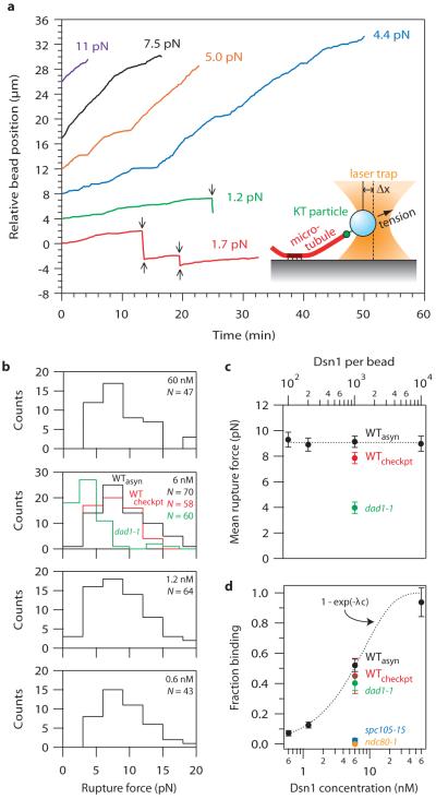
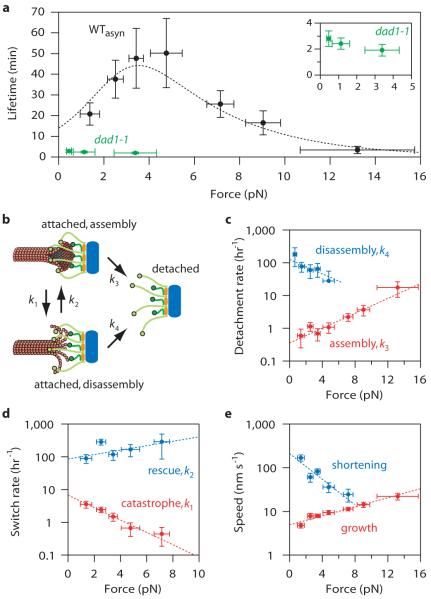
Kinetochores are macromolecular machines that couple chromosomes to dynamic microtubule tips during cell division, thereby generating force to segregate the chromosomes. Accurate segregation depends on selective stabilization of correct 'bi-oriented' kinetochore-microtubule attachments, which come under tension as the result of opposing forces exerted by microtubules. Tension is thought to stabilize these bi-oriented attachments indirectly, by suppressing the destabilizing activity of a kinase, Aurora B. However, a complete mechanistic understanding of the role of tension requires reconstitution of kinetochore-microtubule attachments for biochemical and biophysical analyses in vitro. Here we show that native kinetochore particles retaining the majority of kinetochore proteins can be purified from budding yeast and used to reconstitute dynamic microtubule attachments. Individual kinetochore particles maintain load-bearing associations with assembling and disassembling ends of single microtubules for >30 min, providing a close match to the persistent coupling seen in vivo between budding yeast kinetochores and single microtubules. Moreover, tension increases the lifetimes of the reconstituted attachments directly, through a catch bond-like mechanism that does not require Aurora B. On the basis of these findings, we propose that tension selectively stabilizes proper kinetochore-microtubule attachments in vivo through a combination of direct mechanical stabilization and tension-dependent phosphoregulation.


Figures

Figure 1
Figure 1. Kinetochore particles can be isolated by affinity purification of Dsn1-FLAG
a, Core kinetochore proteins co-purify with Dsn1-FLAG as visualized by silver-stained SDS-PAGE. Asterisk indicates non-specific co-purifying proteins. b, Immunoblot confirms co-association of DNA- and microtubule-binding components with Dsn1-FLAG. c, Mass spectrometry identifies all components of kinetochore subcomplexes except CBF3. Identities, percent sequence coverage, and number of identified peptides of core kinetochore proteins are shown. See Table S1 for all proteins identified by mass spectrometry. d, Eluted kinetochore particles were subjected to S-500 size exclusion chromatography and analyzed by immunoblots. The kinetochore proteins analyzed co-migrate as a complex (Stokes radius ~25 nm). Fig. S3 shows additional fractions.
Figure 2
Figure 2. Purified kinetochore particles bind microtubules in vitro
a, Silver-stained SDS-PAGE of Dsn1-HIS-FLAG kinetochore material from wild-type (WT), ndc80-1, spc105-15 and dad1-1 mutants. Red dots indicate reduced proteins in the mutant preparations (see Fig. S4). Bottom: Anti-FLAG immunoblot against Dsn1-HIS-FLAG. b, Binding of beads prepared with material from indicated strains to taxol-stabilized microtubules (mean ± s.d., from N fields, as indicated). c, Fluorescence images of Cse4-GFP kinetochore particles (green) from wild-type and ndc80-1 strains bound to taxol-stabilized microtubules (red). d, Selected frames from Movie S1 showing movement of Cse4-GFP particles (arrows) driven by the disassembling ends of a microtubule (red; see also, Fig. S7 and Movie S2).
Figure 3
Figure 3. Single kinetochore particles suffice for robust coupling
a, Records of position versus time for native kinetochore-based attachments at indicated tensile loads. Arrows mark transitions (catastrophes and rescues). b, Rupture force distributions for beads prepared with particles at indicated concentrations from wild-type (WTasyn), dad1-1, and checkpoint-activated wild-type cells (WTcheckpt). c, Mean rupture force (± s.d., from N ruptures, indicated in b) versus labeling density, expressed as the Dsn1-HIS-FLAG concentration (bottom scale) and the corresponding Dsn1:bead ratio (top scale). d, Fraction of beads that bound a growing microtubule end (mean ± s.d., N = 11-396). Dotted curve shows Poisson fit (see text and Supplementary Information for details).
Figure 4
Figure 4. Tension stabilizes attachments between kinetochore particles and dynamic microtubules
a, Measured attachment lifetimes for wild-type (WTasyn) and dad1-1 particles. Tension initially prolongs and then shortens lifetimes for wild-type attachments. Dotted curve shows prediction of the two-state model (see text). b, Schematic of two-state model with detachment during assembly and disassembly (rates k3 and k4, respectively), and interconversion between states (k1 and k2). c - e, Measured rates and exponential fits for detachment during assembly (c, red), detachment during disassembly (c, blue), catastrophe (d, red), rescue (d, blue), growth (e, red) and shortening (e, blue). Errors represent (a-d) counting uncertainty (N = 24-65) and (e) s.e.m. (N = 78-360).

Comment in

References

    1. Santaguida S, Musacchio A. The life and miracles of kinetochores. EMBO J. 2009;28:2511–2531. - PMC - PubMed
    1. Cheeseman IM, Desai A. Molecular architecture of the kinetochore-microtubule interface. Nat Rev Mol Cell Biol. 2008;9:33–46. - PubMed
    1. Nicklas RB. The forces that move chromosomes in mitosis. Ann. Rev. Biophys. Biophys. Chem. 1988;17:431–449. - PubMed
    1. Maresca TJ, Salmon ED. Welcome to a new kind of tension: translating kinetochore mechanics into a wait-anaphase signal. J Cell Sci. 2010;123:825–835. - PMC - PubMed
    1. Liu D, Vader G, Vromans MJ, Lampson MA, Lens SM. Sensing chromosome bi-orientation by spatial separation of aurora B kinase from kinetochore substrates. Science. 2009;323:1350–1353. - PMC - PubMed

Methods Additional References

    1. Wigge PA, et al. Analysis of the Saccharomyces spindle pole by matrix-assisted laser desorption/ionization (MALDI) mass spectrometry. J Cell Biol. 1998;141:967–977. - PMC - PubMed
    1. Enquist-Newman M, et al. Dad1p, third component of the Duo1p/Dam1p complex involved in kinetochore function and mitotic spindle integrity. Mol Biol Cell. 2001;12:2601–2613. - PMC - PubMed
    1. Longtine MS, et al. Additional modules for versatile and economical PCR-based gene deletion and modification in Saccharomyces cerevisiae. Yeast. 1998;14:953–961. - PubMed
    1. Gelbart ME, Rechsteiner T, Richmond TJ, Tsukiyama T. Interactions of Isw2 chromatin remodeling complex with nucleosomal arrays: analyses using recombinant yeast histones and immobilized templates. Mol Cell Biol. 2001;21:2098–2106. - PMC - PubMed
    1. Sikorski RS, Hieter P. A system of shuttle vectors and yeast host strains designed for efficient manipulation of DNA in Saccharomyces cerevisiae. Genetics. 1989;122:19–27. - PMC - PubMed

Publication types

MeSH terms

Substances