. 2010 Nov 25;468(7323):580-4.
doi: 10.1038/nature09621.

Nanoscale architecture of integrin-based cell adhesions

Affiliations

Nanoscale architecture of integrin-based cell adhesions

Pakorn Kanchanawong et al. Nature. .

Abstract

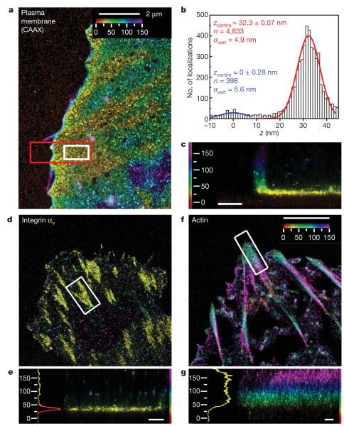
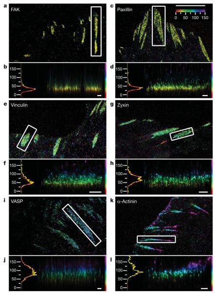
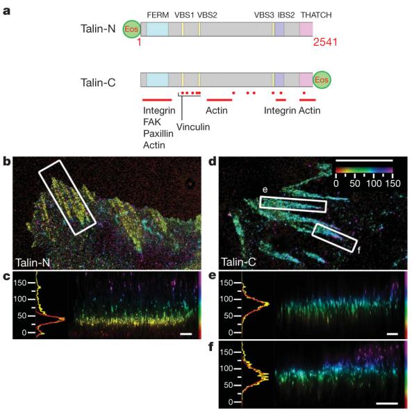
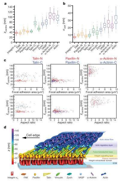
Cell adhesions to the extracellular matrix (ECM) are necessary for morphogenesis, immunity and wound healing. Focal adhesions are multifunctional organelles that mediate cell-ECM adhesion, force transmission, cytoskeletal regulation and signalling. Focal adhesions consist of a complex network of trans-plasma-membrane integrins and cytoplasmic proteins that form a <200-nm plaque linking the ECM to the actin cytoskeleton. The complexity of focal adhesion composition and dynamics implicate an intricate molecular machine. However, focal adhesion molecular architecture remains unknown. Here we used three-dimensional super-resolution fluorescence microscopy (interferometric photoactivated localization microscopy) to map nanoscale protein organization in focal adhesions. Our results reveal that integrins and actin are vertically separated by a ∼40-nm focal adhesion core region consisting of multiple protein-specific strata: a membrane-apposed integrin signalling layer containing integrin cytoplasmic tails, focal adhesion kinase and paxillin; an intermediate force-transduction layer containing talin and vinculin; and an uppermost actin-regulatory layer containing zyxin, vasodilator-stimulated phosphoprotein and α-actinin. By localizing amino- and carboxy-terminally tagged talins, we reveal talin's polarized orientation, indicative of a role in organizing the focal adhesion strata. The composite multilaminar protein architecture provides a molecular blueprint for understanding focal adhesion functions.


Figures

Figure 1
Figure 1. iPALM imaging of a plasma membrane marker, integrin αv and actin
a–c, Plasma membrane marker CAAX–tdEos. a, Top view; b, histogram and Gaussian fits for the z positions (white box in a) of PA-FP molecules (red) and nonspecific fluorescence adsorbed to substrate (blue); c, side view (red box in a). d, e, Integrin αv–tdEos. d, Top view; e, side view (right), histogram and fits (left). f, g, Actin–mEos2. f, Top view; g, side view (right), histograms and fits (left). The vertical distribution of actin is non-Gaussian, so the focal adhesion peak fit is not shown. Colours in a, c–g indicate the vertical (z) coordinate relative to the substrate (z=0nm, red). Scale bars: 500 nm (c, e, g).
Figure 2
Figure 2. Protein stratification of the focal adhesion core
Top view and side view iPALM images of focal adhesions (white boxes, top-view panels) and corresponding z histograms and fits. a, b, FAK–tdEos; c, d, paxillin–tdEos; e, f, vinculin–tdEos; g, h, zyxin–mEos2; i, j, VASP–mEos2; k, l, α-actinin–mEos2. The vertical distribution of α-actinin is non-Gaussian, so the focal adhesion peak fit is not shown. Paxillin and α-actinin shown are C-terminal PA-FP-tagged (N-terminal fusions in Supplementary Figs 21 and 22). Colours: vertical (z) coordinate relative to the substrate (z=0nm, red). Scale bars: 5 μm (a, c, e, g, i, k) and 500 nm (b, d, f, h, j, l).
Figure 3
Figure 3. Talin orientation in focal adhesions
a, Schematic diagram, with important domains and binding sites indicated for Talin PA-FP fusions (FERM, protein 4.1, ezrin, radixin, moesin domain; VBS, vinculin binding sequence; IBS, integrin binding site). Talin-N, N-terminal fusion; Talin-C, C-terminal fusion (Supplementary Table 3). b–f, Top view and side view iPALM images of focal adhesions (white boxes, top-view panels) and corresponding z histograms and fits for talin-N–tdEos (b, c) and talin-C–tdEos (d–f). Colours: vertical (z) coordinate relative to the substrate (z=0nm, red). Scale bars: 5 μm (b, d) and 500 nm (c, e, f).
Figure 4
Figure 4. Nanoscale architecture of focal adhesions
a, b, Peak position (zcentre)(a) and width parameter (σvert)(b) of PA-FP fusions in focal adhesions. Notched boxes, 1st and 3rd quartiles, median and confidence interval; whiskers, 5th and 95th percentiles; +, means, outliers also shown. (See also Supplementary Table 1.) c, zcentre protein positions (nm) versus focal adhesion area (μm2) or aspect ratio for both N- (red) or C- (blue) terminal fusions of talin, paxillin and α-actinin. Each point corresponds to individual focal adhesion measurements. d, Schematic model of focal adhesion molecular architecture, depicting experimentally determined protein positions. Note that the model does not depict protein stoichiometry.

Comment in

  • Cell adhesion: Getting to the core.
    Legg K. Legg K. Nat Rev Mol Cell Biol. 2011 Jan;12(1):4. doi: 10.1038/nrm3030. Epub 2010 Dec 1. Nat Rev Mol Cell Biol. 2011. PMID: 21119700 No abstract available.

Similar articles

Cited by

References

    1. Burridge K, Chrzanowska-Wodnicka M. Focal adhesions, contractility, and signaling. Annu. Rev. Cell Dev. Biol. 1996;12:463–518. - PubMed
    1. Geiger B, Bershadsky A, Pankov R, Yamada KM. Transmembrane crosstalk between the extracellular matrix-cytoskeleton. Nature Rev. Mol. Cell Biol. 2001;2:793–805. - PubMed
    1. Bershadsky AD, Balaban NQ, Geiger B. Adhesion-dependent cell mechanosensitivity. Annu. Rev. Cell Dev. Biol. 2003;19:677–695. - PubMed
    1. Zaidel-Bar R, et al. Functional atlas of the integrin adhesome. Nature Cell Biol. 2007;9:858–867. - PMC - PubMed
    1. Franz CM, Muller DJ. Analyzing focal adhesion structure by atomic force microscopy. J. Cell Sci. 2005;118:5315–5323. - PubMed

Publication types