Review
. 2011 Jan;248(1):181-90.
doi: 10.1007/s00709-010-0239-0. Epub 2010 Nov 24.

Symplasmic transport and phloem loading in gymnosperm leaves

Affiliations
Review

Symplasmic transport and phloem loading in gymnosperm leaves

Johannes Liesche et al. Protoplasma. 2011 Jan.

Abstract

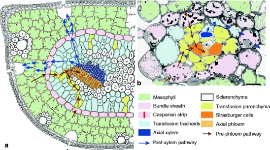
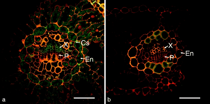
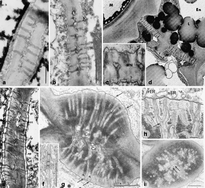
Despite more than 130 years of research, phloem loading is far from being understood in gymnosperms. In part this is due to the special architecture of their leaves. They differ from angiosperm leaves among others by having a transfusion tissue between bundle sheath and the axial vascular elements. This article reviews the somewhat inaccessible and/or neglected literature and identifies the key points for pre-phloem transport and loading of photoassimilates. The pre-phloem pathway of assimilates is structurally characterized by a high number of plasmodesmata between all cell types starting in the mesophyll and continuing via bundle sheath, transfusion parenchyma, Strasburger cells up to the sieve elements. Occurrence of median cavities and branching indicates that primary plasmodesmata get secondarily modified and multiplied during expansion growth. Only functional tests can elucidate whether this symplasmic pathway is indeed continuous for assimilates, and if phloem loading in gymnosperms is comparable with the symplasmic loading mode in many angiosperm trees. In contrast to angiosperms, the bundle sheath has properties of an endodermis and is equipped with Casparian strips or other wall modifications that form a domain border for any apoplasmic transport. It constitutes a key point of control for nutrient transport, where the opposing flow of mineral nutrients and photoassimilates has to be accommodated in each single cell, bringing to mind the principle of a revolving door. The review lists a number of experiments needed to elucidate the mode of phloem loading in gymnosperms.


Figures

Fig. 1
Fig. 1
Illustration of the different cell types and transport pathways in gymnosperm leaves on a schematic drawing of a P. sylvestris needle cross section (a) and an electron micrograph of a Gnetum gnemon fifth class minor vein (b). The pre-phloem pathway (brown arrows) from the mesophyll is symplasmic and crosses bundle sheath, transfusions parenchyma and Strasburger cells before approaching the sieve elements. The post-xylem pathway (blue arrows) in pines starts in transfusion tracheids and has to enter the bundle sheath via the inner tangential wall, since the radial walls are sealed by the suberized Casparian strips (red). From the bundle sheath onwards it is not known to which extent the transpiration stream follows an apoplasmic, symplasmic, or transcellular route towards the sub-stomatal chambers (indicated here by the parallel arrows in a); scale bar 10 μm; a based on Fig. 23 in Münch (1930)
Fig. 2
Fig. 2
Fluorescence micrographs of cross-sections of fixed leaves stained with the component-specific cell wall dyes Berberine hemisulfate (lignin and suberin), Aniline blue (quenching of nonspecific Berberine hemisulfate staining) and Coriphosphine B (pectin) to highlight wall modifications, especially the Casparian strip (Cs) in the Pinus parviflora needle (a) and the suberized bundle sheath of G. biloba (b); En endodermis-like bundle sheath, P phloem, X xylem; scale bars 50 μm
Fig. 3
Fig. 3
Electron micrographs of plasmodesmata at the interfaces between two mesophyll cells (a, Boddi et al. 2002), a mesophyll (left) and an endodermis cell (right) (b, Boddi et al. 2002), two endodermis cells (radial wall) (c, Carde ; d, Gambles and Dengler 1982), a transfusion parenchyma cell (left) and a Strasburger cell (right) (e, Carde 1974), and two Strasburger cells (fi, Glockmann and Kollmann 1996). All contacts in mature cell walls are characterized by branched plasmodesmata and more or less extended median cavities (all but f). Simple plasmodesmata connect young Strasburger cells (f), but get secondarily modified in mature Strasburger cells by the development of dome-shaped wall thickenings that contain complex branched plasmodesmal connections (g)–(i). Neck region at the orifices of these contacts (h). Arrow in d = thickened wall in the center of the pit, in g = neck regions, in h = branching site, in i = bulb-like dilation in cavity, arrowheads in g = expanded sleeve regions, Ca and cc central cavity, En endodermal cell, ep periplasmatic space, L lipid droplet, M mesophyll cell, Mi mitochondria, MN median nodule, pl plasmalemma, rER rough endoplasmatic reticulum, sER smooth endoplasmatic reticulum, tp desmotuble; scale bars 1 μm (a, b, g, f, i), 5 μm (e), 0.1 μm (h)

Similar articles

Cited by

References

    1. Boddi S, Bonzi LM, Calamassi R. Structure and ultrastructure of Pinus halepensis primary needles. Flora. 2002;197(1):10–23.
    1. Burch-Smith TM, Stonebloom S, Xu M, Zambryski PC (2010) Plasmodesmata during development: re-examination of the importance of primary, secondary, and branched plasmodesmata structure versus function. Protoplasma - PMC - PubMed
    1. Canny MJ. Transfusion tissue of pine needles as a site of retrieval of solutes from the transpiration stream. New Phytol. 1993;123(2):227–232. doi: 10.1111/j.1469-8137.1993.tb03730.x. - DOI
    1. Canny MJ. Apoplastic water and solute movement: new rules for an old space. Annu Rev Plant Physiol Plant Mol Biol. 1995;46:215–236. doi: 10.1146/annurev.pp.46.060195.001243. - DOI
    1. Carde JP. Le tissu de transfert (=cellules de Strasburger) dans les aiguilles du pin maritime (Pinus pinaster ait.), I. Étude histologique et infrastructurale du tissu adulte. J Microsc. 1973;17:65–88.

LinkOut - more resources