Review
. 2011 Feb;68(3):369-95.
doi: 10.1007/s00018-010-0580-7. Epub 2010 Nov 24.

Implications of the polymorphism of HLA-G on its function, regulation, evolution and disease association

Affiliations
Review

Implications of the polymorphism of HLA-G on its function, regulation, evolution and disease association

Eduardo A Donadi et al. Cell Mol Life Sci. 2011 Feb.

Abstract

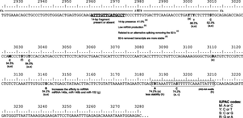
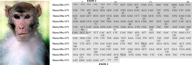
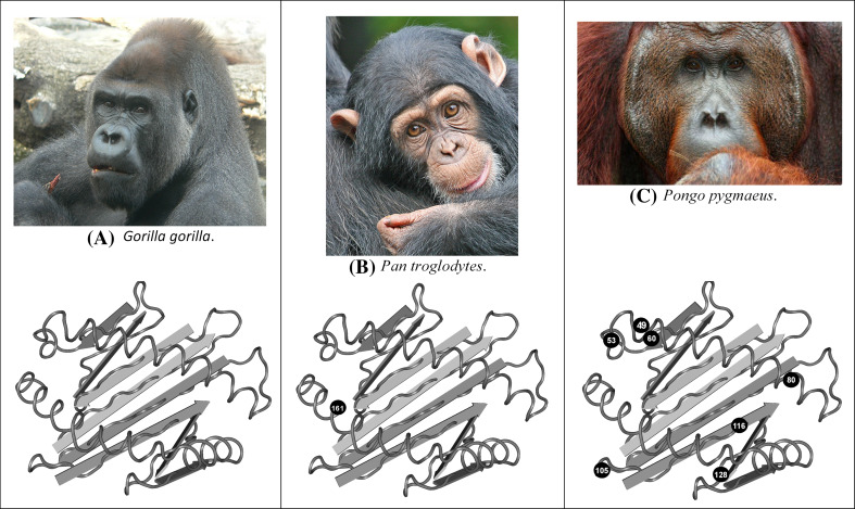
The HLA-G gene displays several peculiarities that are distinct from those of classical HLA class I genes. The unique structure of the HLA-G molecule permits a restricted peptide presentation and allows the modulation of the cells of the immune system. Although polymorphic sites may potentially influence all biological functions of HLA-G, those present at the promoter and 3' untranslated regions have been particularly studied in experimental and pathological conditions. The relatively low polymorphism observed in the MHC-G coding region both in humans and apes may represent a strong selective pressure for invariance, whereas, in regulatory regions several lines of evidence support the role of balancing selection. Since HLA-G has immunomodulatory properties, the understanding of gene regulation and the role of polymorphic sites on gene function may permit an individualized approach for the future use of HLA-G for therapeutic purposes.


Figures

Fig. 1
Fig. 1
Nucleotide sequences, from exon 1 to 4, described for the 44 alleles or haplotypes observed in the coding region of the HLA-G gene (IMGT version 2.28.0, January 2010). Asterisks represent that no official information regarding these single nucleotide polymorphisms was available. Hyphen indicates deletion. Amino acid codes: A alanine, S serine, F phenylalanine, Y tyrosine, T threonine, M methionine, Q glutamine, R arginine, E glutamic acid, P proline, H histidine, G glycine, D aspartic acid, V valine, C cysteine, L leucine, I isoleucine, W tryptophan
Fig. 2
Fig. 2
HLA-G α1, α2 and α3 domains. Variable amino acid positions are indicated in blue circles for each domain (codons). The consensus amino acids are based on the *01:01:01:01 amino acid sequence, and the amino acid exchange for each coding-region allele is given. Probably G*01:13N is not expressed; all domains of the G*01:05N may be expressed. Based on IMGT database version 2.28.0, January 2010. See also [12, 109]
Fig. 3
Fig. 3
Isoforms of HLA-G produced by alternative splicing of the primary mRNA. The HLA-G heavy chain domains (α1, α2, α3, transmembrane portion and cytoplasmic tail) are encoded by the HLA-G gene (chromosome 6), and the light β2-microglobulin molecule is encoded by a gene at chromosome 15. Exon 7 is always spliced out. The final portion of exon 6 and exon 8 is always transcribed, however, never translated due to the presence of a premature stop codon at the beginning of exon 6 (red stop signal), and has been considered as the 3′untranslated region of the gene (3′UTR). The primary transcript may be spliced out into 7 isoforms, HLA-G1 to −G7. HLA-G1 is the full-length HLA-G molecule, HLA-G2 lacks exon 3, HLA-G3 lacks exons 3 and 4, and HLA-G4 lacks exon 4. HLA-G1 to −G4 are membrane-bound molecules due to the presence of the transmembrane and cytoplasmic tail encoded by exons 5 and 6. HLA-G5 is similar to HLA-G1 but retains intron 4, HLA-G6 lacks exon 3 but retains intron 4, and HLA-G7 lacks exon 3 but retains intron 2. HLA-G5 and -G6 are soluble forms due to the presence of intron 4, which contains a premature stop codon at exon 4 (blue stop signal), preventing the translation of the transmembrane and cytoplasmic tail. HLA-G7 is soluble due to the presence of intron 2, which presents a premature stop codon (green stop signal). The G*01:13N allele is probably not expressed due to the presence of a premature stop codon at exon 2 (codon 54). A deletion of a cytosine (ΔC) at exon 3 of the G*01:05N allele changes the reading frame, leading to a stop codon at exon 4
Fig. 4
Fig. 4
Upper panel Comparison of cis-regulatory elements of classical HLA-class I and HLA-G proximal promoter regions (200 base pairs upstream of ATG). Dotted boxes with slashes indicate mutated regulatory elements in the HLA-G promoter. Mutations prevent binding of major classical HLA-class I transacting factors. RFX complex binds to the conserved HLA-G X1 box in electrophoretic mobility shift assay but is not associated to the HLA-G promoter in situ. Lower panel Single nucleotide polymorphisms along the whole HLA-G gene promoter sequence (1.4 kb upstream of ATG) as well as the location of the known regulatory elements
Fig. 5
Fig. 5
Variation sites, described by us and by other authors, observed in the HLA-G 3′ untranslated region, which may influence HLA-G expression. The frequencies of some of these polymorphic sites observed in a highly heterogenous population such as Brazilians are also shown. a Castelli et al. [82]. b Hviid et al. [87]. c Hiby et al. [85]. d Rousseau et al. [70]. e Alvarez et al. [74]. f Unpublished data. This SNP was found in the Brazilian population in only two individuals. g Tan et al. [81]. h Yie et al. [91]
Fig. 6
Fig. 6
New World monkeys: the cotton-top tamarin (Saguinus oedipus, Saoe) MHC-C. Two different MHC-C sequences have been found (GenBank accession numbers AF005217 and AF005218), which cluster with other primates MHC-C. This is represented in a NJ dendrogram. HLA human MHC, Patr chimpanzee, Gogo gorilla, Popy orangutan, Hyla gibbon, Mamu M. mulatta
Fig. 7
Fig. 7
The MHC-G DNA sequence of cotton-top tamarins (Saoe) is more closely related to the primate MHC-E than to the primate MHC-G sequence in the NJ tree (shown), also regarding DNA base percentage similarity (not shown). HLA human MHC, Patr chimpanzee MHC, Gogo gorilla MHC, Popy orangutan MHC, Hyla gibbon MHC, Mamu M. mulatta
Fig. 8
Fig. 8
MHC-G intron 2 nucleotide sequences compared to other MHC sequences (see also [98, 101]. The box indicates the 23-bp deletion observed in all introns 2 from MHC-G sequences. Cotton-top tamarins (Saoe) do not show this typical deletion, and this feature further casts doubts about whether the described cotton-top tamarin’s majority MHC alleles belong to the MHC-G lineage. Otherwise, this deletion may have appeared in the Old World monkey’s lineage. Saoe intron 2 sequence belongs to Saoe-G*02 and -G*04 alleles, which were sequenced in three different monkeys. Identity between residues is indicated by a dash and deletions are denoted by an asterisk
Fig. 9
Fig. 9
Saoe (cotton-top tamarins) MHC-G clusters in relatedness dendrograms with MHC-E of other primates (see Fig. 7)
Fig. 10
Fig. 10
Macaca mulatta. MHC-G exon 2 and exon 3 sequences, showing two stop codon positions at exon 3 (TGA) (five individuals were studied). Macaca fascicularis (four individuals) and Cercopithecus aethiops (five individuals) also showed stop codons at MHC-G exon 3. All of these monkeys belong to the Cercopithecinae family and show one stop codon at the 164 position. Some other Macaca species (43 individuals were studied) show additional stop codons at 118, 133 and 176 exon 3 residues [101]
Fig. 11
Fig. 11
MHC-G molecule (α1, α2 and α3 domains) in primates. a Gorilla gorilla: only one MHC-G allele was found; b Pan troglodytes: one variable position and two different MHC-G alleles were found; and c Pongo pygmaeus: MHC-G alleles found in variable positions in five orangutans. Variable amino acid positions are indicated in circles for each domain cluster (codons)

Similar articles

Cited by

References

    1. Robinson J, Waller MJ, Fail SC, Marsh SG. The IMGT/HLA and IPD databases. Hum Mutat. 2006;27:1192–1199. doi: 10.1002/humu.20406. - DOI - PubMed
    1. Carosella ED, Favier B, Rouas-Freiss N, Moreau P, Lemaoult J. Beyond the increasing complexity of the immunomodulatory HLA-G molecule. Blood. 2008;111:4862–4870. doi: 10.1182/blood-2007-12-127662. - DOI - PubMed
    1. Ishitani A, Sageshima N, Lee N, Dorofeeva N, Hatake K, Marquardt H, Geraghty DE. Protein expression and peptide binding suggest unique and interacting functional roles for HLA-E, F, and G in maternal-placental immune recognition. J Immunol. 2003;171:1376–1384. - PubMed
    1. Diehl M, Munz C, Keilholz W, Stevanovic S, Holmes N, Loke YW, Rammensee HG. Nonclassical HLA-G molecules are classical peptide presenters. Curr Biol. 1996;6:305–314. doi: 10.1016/S0960-9822(02)00481-5. - DOI - PubMed
    1. Lee N, Malacko AR, Ishitani A, Chen MC, Bajorath J, Marquardt H, Geraghty DE. The membrane-bound and soluble forms of HLA-G bind identical sets of endogenous peptides but differ with respect to TAP association. Immunity. 1995;3:591–600. doi: 10.1016/1074-7613(95)90130-2. - DOI - PubMed

Publication types

MeSH terms