

Diseases affecting bone quality: beyond osteoporosis
- PMID: 21107923
- PMCID: PMC3126973
- DOI: 10.1007/s11999-010-1694-9
Diseases affecting bone quality: beyond osteoporosis
Abstract
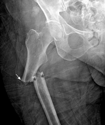
Background: Bone quantity, quality, and turnover contribute to whole bone strength. Although bone mineral density, or bone quantity, is associated with increased fracture risk, less is known about bone quality. Various conditions, including disorders of mineral homeostasis, disorders in bone remodeling, collagen disorders, and drugs, affect bone quality.
Questions/purposes: The objectives of this review are to (1) identify the conditions and diseases that could adversely affect bone quality besides osteoporosis, and (2) evaluate how these conditions influence bone quality.
Methods: We searched PubMed using the keywords "causes" combined with "secondary osteoporosis" or "fragility fracture." After identifying 20 disorders/conditions, we subsequently searched each condition to evaluate its effect on bone quality.
Results: Many disorders or conditions have an effect on bone metabolism, leading to fragility fractures. These disorders include abnormalities that disrupt mineral homeostasis, lead to an alteration of the mineralization process, and ultimately reduce bone strength. The balance between bone formation and resorption is also essential to prevent microdamage accumulation and maintain proper material and structural integrity of the bone. As a result, diseases that alter the bone turnover process lead to a reduction of bone strength. Because Type I collagen is the most abundant protein found in bone, defects in Type I collagen can result in alterations of material property, ultimately leading to fragility fractures. Additionally, some medications can adversely affect bone.
Conclusions: Recognizing these conditions and diseases and understanding their etiology and pathogenesis is crucial for patient care and maintaining overall bone health.
Figures
Similar articles
-
Factors affecting bone strength other than osteoporosis.Aging Clin Exp Res. 2013 Oct;25 Suppl 1:S9-11. doi: 10.1007/s40520-013-0098-6. Epub 2013 Sep 18. Aging Clin Exp Res. 2013. PMID: 24046057 Review.
-
The actions of parathyroid hormone on bone: relation to bone remodeling and turnover, calcium homeostasis, and metabolic bone disease. Part IV of IV parts: The state of the bones in uremic hyperaparathyroidism--the mechanisms of skeletal resistance to PTH in renal failure and pseudohypoparathyroidism and the role of PTH in osteoporosis, osteopetrosis, and osteofluorosis.Metabolism. 1976 Oct;25(10):1157-88. doi: 10.1016/0026-0495(76)90024-x. Metabolism. 1976. PMID: 787723 Review.
-
Insights into material and structural basis of bone fragility from diseases associated with fractures: how determinants of the biomechanical properties of bone are compromised by disease.Endocr Rev. 2007 Apr;28(2):151-64. doi: 10.1210/er.2006-0029. Epub 2006 Dec 19. Endocr Rev. 2007. PMID: 17200084 Review.
-
Bone quality and osteoporosis therapy.Arq Bras Endocrinol Metabol. 2010 Mar;54(2):186-99. doi: 10.1590/s0004-27302010000200015. Arq Bras Endocrinol Metabol. 2010. PMID: 20485908 Review.
-
Important determinants of bone strength: beyond bone mineral density.J Clin Rheumatol. 2006 Apr;12(2):70-7. doi: 10.1097/01.rhu.0000208612.33819.8c. J Clin Rheumatol. 2006. PMID: 16601540 Review.
Cited by
-
Inhibition of cysteine protease disturbs the topological relationship between bone resorption and formation in vitro.J Bone Miner Metab. 2024 Mar;42(2):166-184. doi: 10.1007/s00774-023-01489-w. Epub 2024 Feb 20. J Bone Miner Metab. 2024. PMID: 38376670 Free PMC article.
-
The impact of medication on osseointegration and implant anchorage in bone determined using removal torque-A review.Heliyon. 2022 Oct 3;8(10):e10844. doi: 10.1016/j.heliyon.2022.e10844. eCollection 2022 Oct. Heliyon. 2022. PMID: 36276721 Free PMC article.
-
Higher risk of 2-year cup revision of ceramic-on-ceramic versus ceramic-on-polyethylene bearing: analysis of 33,454 primary press-fit total hip arthroplasties registered in the Dutch Arthroplasty Register (LROI).Hip Int. 2023 Mar;33(2):280-287. doi: 10.1177/11207000211064975. Epub 2022 Jan 2. Hip Int. 2023. PMID: 34974763 Free PMC article.
-
Osteoporosis Associated with Chronic Obstructive Pulmonary Disease.J Bone Metab. 2016 Aug;23(3):111-20. doi: 10.11005/jbm.2016.23.3.111. Epub 2016 Aug 31. J Bone Metab. 2016. PMID: 27622174 Free PMC article. Review.
-
Bone Quality in Relation to HIV and Antiretroviral Drugs.Curr HIV/AIDS Rep. 2022 Oct;19(5):312-327. doi: 10.1007/s11904-022-00613-1. Epub 2022 Jun 20. Curr HIV/AIDS Rep. 2022. PMID: 35726043 Free PMC article. Review.
References
-
- Abbasi AA, Rudman D, Wilson CR, Drinka PJ, Basu SN, Mattson DE, Richardson TJ. Observations on nursing home residents with a history of hip fracture. Am J Med Sci. 1995;310:229–234. - PubMed
-
- Alman BA, Goldberg MJ. Metabolic and endocrine abnormalities. In: Lovell WW, Winter RB, Morrissy RT, Weinstein SL, editors. Lovell and Winter’s Pediatric Orthopaedics. 6. Philadelphia: Lippincott Williams & Wilkins; 2006. pp. 167–203.
-
- Amir E, Simmons CE, Freedman OC, Dranitsaris G, Cole DE, Vieth R, Ooi WS, Clemons M. A phase 2 trial exploring the effects of high-dose (10, 000 IU/day) vitamin D(3) in breast cancer patients with bone metastases. Cancer. 2010;116:284–291. - PubMed
-
- Baron R, Rawadi G. Targeting the Wnt/beta-catenin pathway to regulate bone formation in the adult skeleton. Endocrinology. 2007;6:2635–2643. - PubMed
Publication types
MeSH terms
Substances
LinkOut - more resources
Full Text Sources
Other Literature Sources
Medical
Research Materials
