. 2011 May;20(5):706-12.
doi: 10.1007/s00586-010-1626-0. Epub 2010 Nov 25.

Lumbopelvic alignment on standing lateral radiograph of adult volunteers and the classification in the sagittal alignment of lumbar spine

Affiliations

Lumbopelvic alignment on standing lateral radiograph of adult volunteers and the classification in the sagittal alignment of lumbar spine

Pongsthorn Chanplakorn et al. Eur Spine J. 2011 May.

Abstract

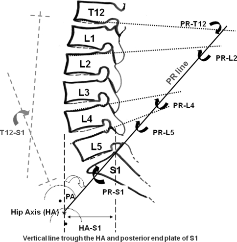
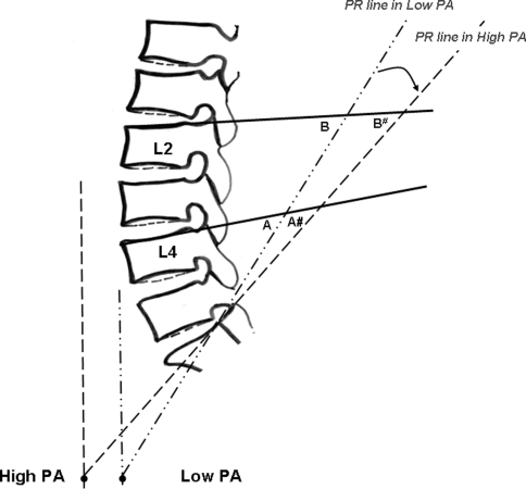
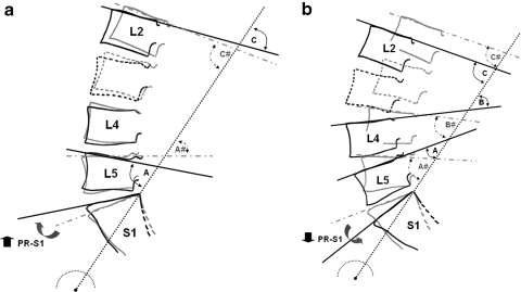
The analysis of the sagittal balance is important for the understanding of the lumbopelvic biomechanics. Results from previous studies documented the correlation between sacro-pelvic orientation and lumbar lordosis and a uniqueness of spino-pelvic alignment in an individual person. This study was subjected to determine the lumbopelvic orientation using pelvic radius measurement technique. The standing lateral radiographs in a standardized standing position were taken from 100 healthy volunteers. The measurements which included hip axis (HA), pelvic radius (PR), pelvic angle (PA), pelvic morphology (PR-S1), sacral translation distance (HA-S1), total lumbosacral lordosis (T12-S1), total lumbopelvic lordosis (PR-T12) and regional lumbopelvic lordosis angles (PR-L2, PR-L4 and PR-L5) were carried out with two independent observers. The relationships between the parameters were as follows. PR-S1 demonstrated positive correlation to regional lumbopelvic lordosis and revealed negative correlation to T12-S1. PA showed negative correlation to PR-S1 and regional lumbopelvic lordosis, but revealed positive correlation to HA-S1. T12-S1 was significantly increased when PR-S1 was lesser than average (35°-45°) and was significantly decreased when PR-S1 was above the average. PR-L4 and PR-L5 were significantly reduced when PR-S1 was smaller than average and only PR-L5 was significantly increased when PR-S1 was above the average. In conclusion, this present study supports that lumbar spine and pelvis work together in order to maintain lumbopelvic balance.


Figures

Fig. 1
Fig. 1
Positioning technique for standing lateral radiograph of the spine and pelvis. The radiographs were taken with 72-inch-long distance from the X-ray source
Fig. 2
Fig. 2
Line drawing showing pelvic radius (PR line) and the pelvic radius measurement technique used in this study. List of the nomenclature with their abbreviation and description is outlined in Table 1. Black dashed lines demonstrate the vertical line trough HA and posterior superior corner of S1. Gray dashed lines show T12-S1 measurement. Arrows indicate the angles of representation
Fig. 3
Fig. 3
Drawing demonstrates the relationship between PA angle and pelvic radius measurements on lumbar spines (PR-L2, PR-L4 and PR-L5). The black dots represent the HA in high and low PA angles. The PR line in low PA angle is indicated by dash dotted lines and the dashed line is the High PA angle. The PR line was rearward projected as the PA angle was higher as indicated by curve arrow. The regional lumbar lordosis angles were then measured and decrease value even without the change of lumbar lordosis (PR-L4: A > A# and PR-L2: B > B#)
Fig. 4
Fig. 4
The drawings demonstrate the lumbar sagittal alignment which was altered by the change of PR-S1 angle (indicated by black arrows), high PR-S1 angle (a) and low PR-S1 angle (b). Gray line shows the lumbar alignment in average PR-S1. See text for the details. The PR-L2 (C and C#) remained unchanged. In high PR-S1 (a) PR-L5 (A) was higher than that of the average PR-S1 (A#). The lumbar spine was then flat. In low PR-S1 (b) PR-L5 (A) and PR-L4 (B) was less than those of the average PR-S1 (A# and B#, respectively). Lumbar lordosis was increased at lower lumbar segments and the lumbar spine appeared to move forward

Similar articles

Cited by

References

    1. Kobayashi T, Atsuta Y, Matsuno T, Takeda N. A longitudinal study of congruent sagittal spinal alignment in as adult cohort. Spine. 2004;29:671–676. doi: 10.1097/01.BRS.0000115127.51758.A2. - DOI - PubMed
    1. Barrey C, Jund J, Noseda O, Roussouly P. Sagittal balance of the pelvis-spine complex and lumbar degenerative diseases. A comparative study about 85 cases. Eur Spine J. 2007;16:1459–1467. doi: 10.1007/s00586-006-0294-6. - DOI - PMC - PubMed
    1. Legaye J, Duval-Beaupère G, Hecquet J, Marty C. Pelvic incidence: a fundamental pelvic parameter for the three dimensional regulation of spinal sagittal curves. Eur Spine J. 1998;7:99–103. doi: 10.1007/s005860050038. - DOI - PMC - PubMed
    1. Vaz G, Roussouly P, Berthonnaud E, Dimnet J. Sagittal morphology and equilibrium of pelvis and spine. Eur Spine J. 2002;11:80–87. doi: 10.1007/s005860000224. - DOI - PMC - PubMed
    1. Mac-Thiong JM, Berthonnaud E, Dimar JR, Betz RR, Labelle H. Sagittal Alignment of the Spine and Pelvis During Growth. Spine. 2004;29:1642–1647. doi: 10.1097/01.BRS.0000132312.78469.7B. - DOI - PubMed

MeSH terms