

Simultaneous optical coherence tomography and laser induced fluorescence imaging in rat model of ovarian carcinogenesis
- PMID: 21108515
- PMCID: PMC3040967
- DOI: 10.4161/cbt.10.5.12531
Simultaneous optical coherence tomography and laser induced fluorescence imaging in rat model of ovarian carcinogenesis
Abstract
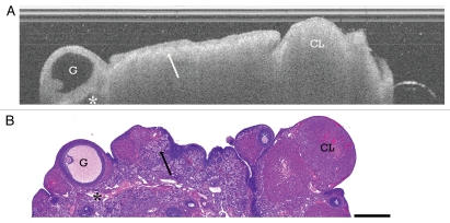
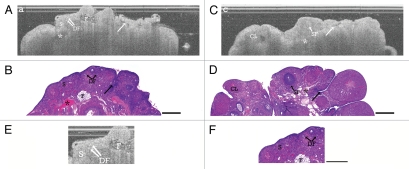
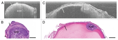
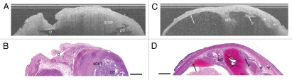
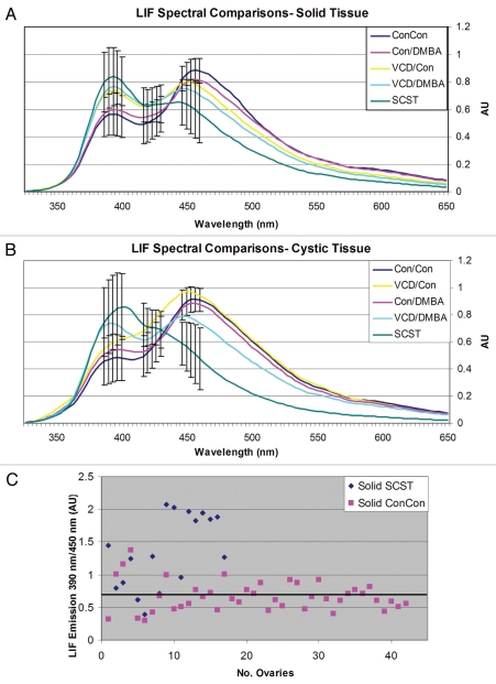
Determining if an ovarian mass is benign or malignant is an ongoing clinical challenge. The development of reliable animal models provides means to evaluate new diagnostic tools to more accurately determine if an ovary has benign or malignant features. Although sex cord-stromal tumors (SCST) account for 0.1–0.5% of ovarian malignancies, they have similar appearances to more aggressive epithelial cancers and can serve as a prototype for developing better diagnostic methods for ovarian cancer. Optical coherence tomography (OCT) and laser-induced fluorescence (LIF) spectroscopy are non-destructive optical imaging modalities. OCT provides architectural cross-sectional images at near histological resolutions and LIF provides biochemical information. We utilize combined OCT-LIF to image ovaries in post-menopausal ovarian carcinogenesis rat models, evaluating normal cyclic, acyclic and neoplastic ovaries. Eighty-three female Fisher rats were exposed to combinations of control sesame oil, 4-vinyl cyclohexene diepoxide (VCD) to induce ovarian failure,and/or 7,12-dimethylbenz[a]anthracene (DMBA) to induce carcinogenesis. Three or five months post-treatment, 162 ovaries were harvested and imaged with OCT-LIF: 40 cyclic, 105 acyclic and 17 SCST. OCT identified various follicle stages,corpora lutea (CL), CL remnants, epithelial invaginations/inclusions and allowed for characterization of both cystic and solid SCST. Signal attenuation comparisons between CL and solid SCST revealed statistically significant increases in attenuation among CL. LIF characterized spectral differences in cyclic, acyclic and neoplastic ovaries attributed to collagen, NADH/FAD and hemoglobin absorption. We present combined OCT-LIF imaging in a rat ovarian carcinogenesis model, providing preliminary criteria for normal cyclic, acyclic and SCST ovaries which support the potential of OCT-LIF for ovarian imaging.
Figures
Similar articles
-
Dual modality imaging of a novel rat model of ovarian carcinogenesis.J Biomed Opt. 2006 Jul-Aug;11(4):041123. doi: 10.1117/1.2236298. J Biomed Opt. 2006. PMID: 16965151
-
Ovarian neoplasm development by 7,12-dimethylbenz[a]anthracene (DMBA) in a chemically-induced rat model of ovarian failure.Gynecol Oncol. 2009 Mar;112(3):610-5. doi: 10.1016/j.ygyno.2008.12.013. Epub 2009 Jan 16. Gynecol Oncol. 2009. PMID: 19150572 Free PMC article.
-
7,12-dimethylbenz[a]anthracene induces sertoli-leydig-cell tumors in the follicle-depleted ovaries of mice treated with 4-vinylcyclohexene diepoxide.Comp Med. 2010 Feb;60(1):10-7. Comp Med. 2010. PMID: 20158943 Free PMC article.
-
An overview of optical coherence tomography for ovarian tissue imaging and characterization.Wiley Interdiscip Rev Nanomed Nanobiotechnol. 2015 Jan-Feb;7(1):1-16. doi: 10.1002/wnan.1306. Epub 2014 Oct 20. Wiley Interdiscip Rev Nanomed Nanobiotechnol. 2015. PMID: 25329515 Free PMC article. Review.
-
Comprehensive review of imaging features of sex cord-stromal tumors of the ovary.Abdom Radiol (NY). 2021 Apr;46(4):1519-1529. doi: 10.1007/s00261-021-02998-w. Epub 2021 Mar 16. Abdom Radiol (NY). 2021. PMID: 33725145 Review.
Cited by
-
Analysis of second-harmonic-generation microscopy in a mouse model of ovarian carcinoma.J Biomed Opt. 2012 Jul;17(7):076002. doi: 10.1117/1.JBO.17.7.076002. J Biomed Opt. 2012. PMID: 22894485 Free PMC article.
-
Two-photon excited fluorescence imaging of endogenous contrast in a mouse model of ovarian cancer.Lasers Surg Med. 2013 Mar;45(3):155-66. doi: 10.1002/lsm.22115. Epub 2013 Jan 29. Lasers Surg Med. 2013. PMID: 23362124 Free PMC article.
-
Optical Resolution Photoacoustic Microscopy of Ovary and Fallopian Tube.Sci Rep. 2019 Oct 4;9(1):14306. doi: 10.1038/s41598-019-50743-7. Sci Rep. 2019. PMID: 31586106 Free PMC article.
-
In vivo molecular imaging of colorectal cancer using quantum dots targeted to vascular endothelial growth factor receptor 2 and optical coherence tomography/laser-induced fluorescence dual-modality imaging.J Biomed Opt. 2015;20(9):096015. doi: 10.1117/1.JBO.20.9.096015. J Biomed Opt. 2015. PMID: 26397238 Free PMC article.
-
Label-Free Optical Metabolic Imaging in Cells and Tissues.Annu Rev Biomed Eng. 2023 Jun 8;25:413-443. doi: 10.1146/annurev-bioeng-071516-044730. Epub 2023 Apr 27. Annu Rev Biomed Eng. 2023. PMID: 37104650 Free PMC article. Review.
References
-
- Cancer Facts and Figures. American Cancer Society; 2008.
-
- Kao SW, Sipes IG, Hoyer PB. Early effects of ovotoxicity induced by 4-vinylcyclohexene diepoxide in rats and mice. Reprod Toxicol. 1999;13:67–75. - PubMed
-
- Springer LN, McAsey ME, Flaws JA, Tilly JL, Sipes IG, Hoyer PB. Involvement of apoptosis in 4-vinylcyclohexene diepoxide-induced ovotoxicity in rats. Toxicol Appl Pharmacol. 1996;139:394–401. - PubMed
-
- Hu X, Flaws JA, Sipes IG, Hoyer PB. Activation of mitogen-activated protein kinases and AP-1 transcription factor in ovotoxicity induced by 4-vinylcyclohexene diepoxide in rats. Biol Reprod. 2002;67:718–724. - PubMed
Publication types
MeSH terms
Substances
Grants and funding
LinkOut - more resources
Full Text Sources
Medical