

Homozygous null mutations in ZMPSTE24 in restrictive dermopathy: evidence of genetic heterogeneity
- PMID: 21108632
- PMCID: PMC3117019
- DOI: 10.1111/j.1399-0004.2010.01580.x
Homozygous null mutations in ZMPSTE24 in restrictive dermopathy: evidence of genetic heterogeneity
Abstract
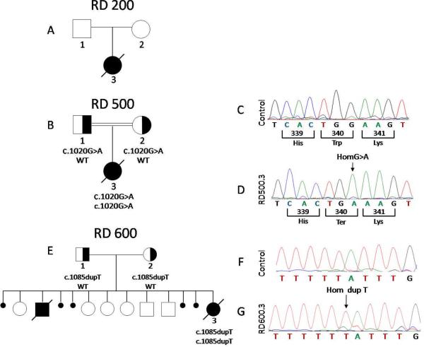
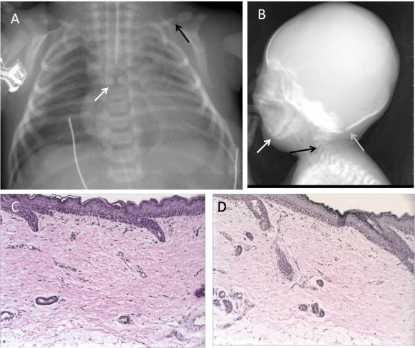
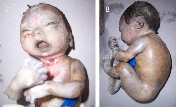
Restrictive dermopathy (RD) results in stillbirth or early neonatal death. RD is characterized by prematurity, intrauterine growth retardation, fixed facial expression, micrognathia, mouth in the 'o' position, rigid and tense skin with erosions and denudations and multiple joint contractures. Nearly all 25 previously reported neonates with RD had homozygous or compound heterozygous null mutations in the ZMPSTE24 gene. Here, we report three new cases of RD; all died within 3 weeks of birth. One of them had a previously reported homozygous c.1085dupT (p.Leu362PhefsX19) mutation, the second case had a novel homozygous c.1020G>A (p.Trp340X) null mutation in ZMPSTE24, but the third case, a stillborn with features of RD except for the presence of tapering rather than rounded, bulbous digits, harbored no disease-causing mutations in LMNA or ZMPSTE24. In the newborn with a novel ZMPSTE24 mutation, unique features included butterfly-shaped thoracic 5 vertebra and the bulbous appearance of the distal clavicles. Skin biopsies from both the stillborn fetus and the newborn with c.1020G>A ZMPSTE24 mutation showed absence of elastic fibers throughout the dermis. This report provides evidence of genetic heterogeneity among RD and concludes that there may be an additional locus for RD which remains to be identified.
© 2010 John Wiley & Sons A/S.
Figures
Similar articles
-
New ZMPSTE24 (FACE1) mutations in patients affected with restrictive dermopathy or related progeroid syndromes and mutation update.Eur J Hum Genet. 2014 Aug;22(8):1002-11. doi: 10.1038/ejhg.2013.258. Epub 2013 Oct 30. Eur J Hum Genet. 2014. PMID: 24169522 Free PMC article.
-
Frame shift mutations of the ZMPSTE24 gene in two siblings with restrictive dermopathy.Clin Dysmorphol. 2016 Jan;25(1):7-11. doi: 10.1097/MCD.0000000000000100. Clin Dysmorphol. 2016. PMID: 26379196
-
Lamin A and ZMPSTE24 (FACE-1) defects cause nuclear disorganization and identify restrictive dermopathy as a lethal neonatal laminopathy.Hum Mol Genet. 2004 Oct 15;13(20):2493-503. doi: 10.1093/hmg/ddh265. Epub 2004 Aug 18. Hum Mol Genet. 2004. PMID: 15317753
-
Restrictive dermopathy--a lethal congenital laminopathy. Case report and review of the literature.Eur J Pediatr. 2009 Aug;168(8):1007-12. doi: 10.1007/s00431-008-0868-x. Epub 2008 Nov 20. Eur J Pediatr. 2009. PMID: 19020898 Review.
-
Restrictive dermopathy: Three new patients with ZMPSTE24 mutations and a review of the literature.Pediatr Dermatol. 2021 Nov;38(6):1535-1540. doi: 10.1111/pde.14822. Epub 2021 Oct 14. Pediatr Dermatol. 2021. PMID: 34647350 Review.
Cited by
-
Phenotypic heterogeneity of ZMPSTE24 deficiency.Am J Med Genet A. 2018 May;176(5):1175-1179. doi: 10.1002/ajmg.a.38493. Epub 2018 Jan 17. Am J Med Genet A. 2018. PMID: 29341437 Free PMC article.
-
Progeroid syndrome patients with ZMPSTE24 deficiency could benefit when treated with rapamycin and dimethylsulfoxide.Cold Spring Harb Mol Case Stud. 2017 Jan;3(1):a001339. doi: 10.1101/mcs.a001339. Cold Spring Harb Mol Case Stud. 2017. PMID: 28050601 Free PMC article.
-
Genomic instability and DNA replication defects in progeroid syndromes.Nucleus. 2018 Dec 31;9(1):368-379. doi: 10.1080/19491034.2018.1476793. Epub 2018 Jun 23. Nucleus. 2018. PMID: 29936894 Free PMC article. Review.
-
New ZMPSTE24 (FACE1) mutations in patients affected with restrictive dermopathy or related progeroid syndromes and mutation update.Eur J Hum Genet. 2014 Aug;22(8):1002-11. doi: 10.1038/ejhg.2013.258. Epub 2013 Oct 30. Eur J Hum Genet. 2014. PMID: 24169522 Free PMC article.
-
Nuclear envelopathies: a complex LINC between nuclear envelope and pathology.Orphanet J Rare Dis. 2017 Aug 30;12(1):147. doi: 10.1186/s13023-017-0698-x. Orphanet J Rare Dis. 2017. PMID: 28854936 Free PMC article. Review.
References
-
- Kariminejad A, Goodarzi P, Thanh Huong le T, et al. Restrictive dermopathy. Molecular diagnosis of restrictive dermopathy in a stillborn fetus from a consanguineous Iranian family. Saudi Med J. 2009;30:150–153. - PubMed
-
- Jagadeesh S, Bhat L, Suresh I, et al. Prenatal diagnosis of restrictive dermopathy. Indian Pediatr. 2009;46:349–351. - PubMed
-
- Morais P, Magina S, Ribeiro Mdo C, et al. Restrictive dermopathy--a lethal congenital laminopathy. Case report and review of the literature. Eur J Pediatr. 2009;168:1007–1012. - PubMed
-
- Navarro CL, Cadinanos J, De Sandre-Giovannoli A, et al. Loss of ZMPSTE24 (FACE-1) causes autosomal recessive restrictive dermopathy and accumulation of Lamin A precursors. Hum Mol Genet. 2005;14:1503–1513. - PubMed
Publication types
MeSH terms
Substances
Supplementary concepts
Grants and funding
LinkOut - more resources
Full Text Sources
Miscellaneous
