. 2011 Apr;10(2):263-76.
doi: 10.1111/j.1474-9726.2010.00660.x. Epub 2010 Dec 29.

Age-related alterations in the dynamic behavior of microglia

Affiliations

Age-related alterations in the dynamic behavior of microglia

Mausam R Damani et al. Aging Cell. 2011 Apr.

Abstract

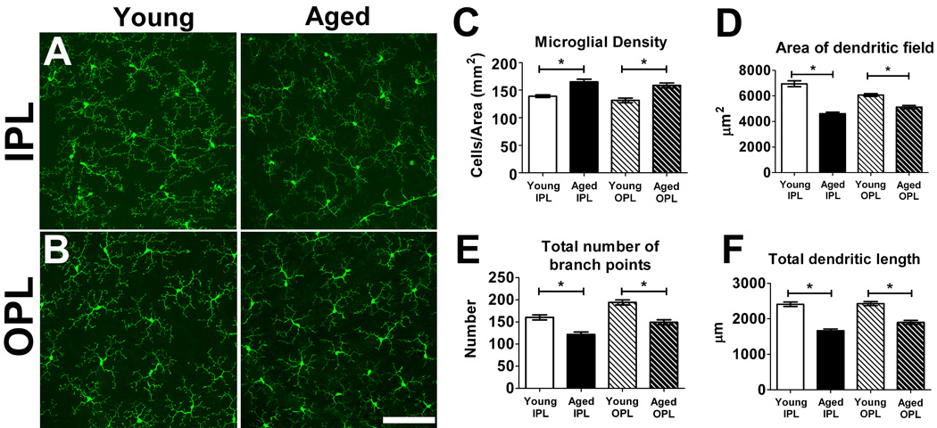
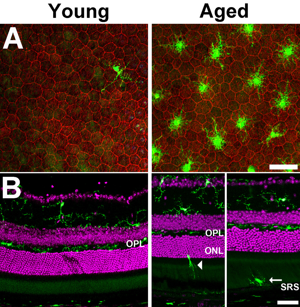
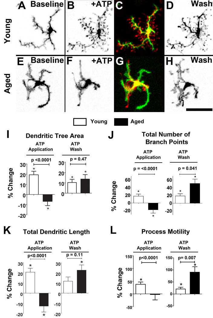
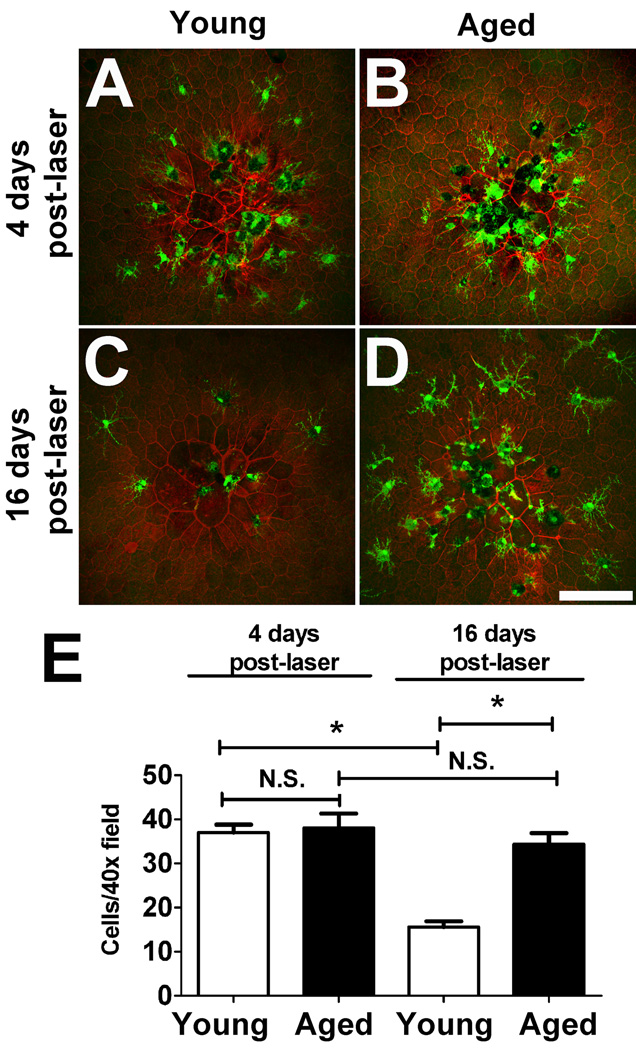
Microglia, the primary resident immune cells of the central nervous system (CNS), exhibit dynamic behavior involving rapid process motility and cellular migration that is thought to underlie key functions of immune surveillance and tissue repair. Although age-related changes in microglial activation have been implicated in the pathogenesis of neurodegenerative diseases of aging, how dynamic behavior in microglia is influenced by aging is not fully understood. In this study, we employed live imaging of retinal microglia in situ to compare microglial morphology and behavioral dynamics in young and aged animals. We found that aged microglia in the resting state have significantly smaller and less branched dendritic arbors, and also slower process motilities, which probably compromise their ability to survey and interact with their environment continuously. We also found that dynamic microglial responses to injury were age-dependent. While young microglia responded to extracellular ATP, an injury-associated signal, by increasing their motility and becoming more ramified, aged microglia exhibited a contrary response, becoming less dynamic and ramified. In response to laser-induced focal tissue injury, aged microglia demonstrated slower acute responses with lower rates of process motility and cellular migration compared with young microglia. Interestingly, the longer term response of disaggregation from the injury site was retarded in aged microglia, indicating that senescent microglial responses, while slower to initiate, are more sustained. Together, these altered features of microglial behavior at rest and following injury reveal an age-dependent dysregulation of immune response in the CNS that may illuminate microglial contributions to age-related neuroinflammatory degeneration.


Figures

Figure 1
Figure 1. Morphology and distribution of retinal microglia in young (2–3 months old) and aged (18–24 months old) CX3CR1+/GFP mice
Resting retinal microglia, as visualized in retinal flat-mount preparations, possess small somata and ramified morphologies that were regularly distributed in the vitreal half of the retina with processes located in the inner plexiform layer (IPL) (A) and the outer plexiform layer (OPL) (B). Qualitatively similar microglial distributions and morphologies were observed in both young (left panels) and aged (right panels) retinas. Scale bar = 100 µm. (C) Densities of microglial somata in both IPL and OPL layers of young and aged retinas were measured in the mid-peripheral retina, showing increased microglial cell densities in the IPL and the OPL in aged compared to young retina (n = 24 retina from 6 young and 6 aged animals). Analyses of morphological parameters of area of dendritic field (D), total number of branch points (E), and total dendritic length of individual microglia (F), revealed that aged microglia have a smaller and less branched dendritic morphologies compared to young microglia in both the IPL and the OPL layers (n = 255 cells from 6 young animals and 205 cells from 6 aged animals). (Asterisks indicate comparisons for which p<0.05)
Figure 2
Figure 2. Translocation of microglia to the outer retina with increasing age
(A) The distribution of retinal microglia in the subretinal space, as visualized in RPE-sclerochoroidal wholemounts, varied between young (left) and aged (right) animals. In young animals, only an occasional microglia cell (green) could be located on the apical surface of RPE cells in the subretinal space (RPE cellular outlines stained with phalloidin, red). In aged animals, significantly greater numbers of microglia accumulated in the subretinal space above RPE cells. These subretinal microglia in the aged retina also had a less ramified morphology with shorter, less branched processes. Scale bar = 50 µm. (B) Changes in microglial distribution with age were also evident in vibratome cross-sections. Retinal microglia in young animals (left) were predominantly confined to the inner retina up to and including the outer plexiform layer (OPL), while in aged animals (right) , retinal microglia could be observed traversing the outer retina in the outer nuclear layer (ONL) (arrowhead) and the subretinal space (SRS)(arrow). Scale bar = 50 µm.
Figure 3
Figure 3. Dynamic process motility in retinal microglia in young and aged CX3CR1+/GFP mice
(A) Representative time-lapse recording of a retinal microglial cell from a young animal, demonstrating dynamic structural plasticity in ramified process. Higher magnification images (inset) of an individual process, taken 80s apart, demonstrate continuous process extensions, retractions, additions, and eliminations. A colorized subtraction image of time-lapse images highlights simultaneous process additions and extensions (in green) and process retractions and eliminations (in red) across a 10-min interval in a representative young retinal microglia cell (B). Positive and negative structural changes were balanced across the dendritic arbor to maintain stable dendritic size, complexity, and symmetry. In aged animals, a qualitatively similar pattern of structural changes in the processes of retinal microglia were found (C, D). (E) Quantitative comparison of process motility in individual retinal microglia located in the inner plexiform layer (IPL) and outer plexiform layer (OPL) of young (n = 42 cells from 4 animals) and aged (n = 49 cells from 4 animals) mice however revealed that mean process motilities were significantly greater in microglia located in the IPL compared to those located in the OPL for both young and aged retina. Comparing young with aged microglia, process motility in aged microglia in the IPL was slightly but not significantly lower, while those in the OPL were significantly lower than their counterparts in the young retina. Scale bars = 50 µm.
Figure 4
Figure 4. Age-dependent changes in microglial morphology and process motility in response to exogenous ATP
Microglia from young animals responded rapidly to bath application of ATP (1 mM) by increasing from their baseline morphological structure (A) by the extension of existing processes and the addition of new processes (B). A colorized subtraction image of time-lapse images across a 10-minute interval in a representative retinal microglial cell demonstrates a predominant addition of new processes and elongation of existing processes (coded in red) (C). Upon washout of ATP, microglia from young retina exhibited further increases in their morphological structure (D). Conversely, microglia from aged animals responded to bath application of ATP (1 mM) by a reducing their morphological structure (E to F), so that a colorized subtraction image primarily demonstrates process retraction (coded in green) (G). Upon washout of ATP, aged microglia, like young microglia, further increased their morphological structure (H). Scale bars = 50 µm. Quantitative analysis of changes in morphological parameters upon the addition and wash-out of ATP are compared in young (white bars) and aged microglia (black bars). Percentage changes in parameters during ATP application were relative to those under baseline conditions, while changes during ATP wash were relative to those during ATP application. Morphological parameters include: dendritic tree area (I), total number of branching points in a single cell (J), and total dendritic length (K). (L) Quantitative analysis of process motility (i.e. rate of movement of processes of individual cells) revealed that while young microglia increased their process motility in response to ATP application, aged microglia demonstrated no significant change. On ATP washout, both young and aged microglia increased their process motility, with aged microglia demonstrating a significantly larger increase (asterisks * indicate significant (p<0.05) differences from baseline values). The number of animals analyzed for (I)–(L) were: young , n = 58 cells from 6 animals; aged, n = 50 cells from 5 animals.
Figure 5
Figure 5. Age-dependent changes in microglial process motility and migratory velocity in response to focal laser injury ex vivo
(A) Migration and polarization of retinal microglia 1.5 hours after the application of focal laser injury (indicated by red spot) in a retinal explant from a young mouse. Microglia polarized their processes toward the site of laser injury and migrated to cluster around the injury site. Scale bar = 100 µm. (B) Quantification of microglial process motility in young and aged retina under baseline condition and following application of focal laser injury. Whereas microglia in young retina significantly increased process motility after laser injury, microglia from aged retina failed to change process motility. (C, D) Microglia in young and aged retina after laser injury acquired a migratory capacity, extending their processes towards the site of injury and directing their migration in that same direction. Time-lapse images taken 10 minutes apart show a similar nature of microglia migration. Superposition of colored images (right) demonstrate displacement of the cell from the start of the recording (red, at t=0) to the end of the recording (green, t=40 min). Scale bar = 50 µm. Migration velocities following laser injury were compared between microglia from young and aged animals. For both mean migratory velocity (E) and maximum instantaneous velocity (F), young microglia exhibited significantly higher values than aged microglia, indicating an age-dependence in post-injury migratory response. Asterisks * indicate significant (p<0.05) differences from baseline values, N.S indicates comparisons for which p >0.05. The number of animals analyzed were: young , n = 74 cells from 7 animals; aged, n = 50 cells from 5 animals.
Figure 6
Figure 6. Age-dependent changes in microglia aggregation and disaggregation following focal laser injury in vivo
Four days following focal laser retinal injury in vivo, microglia in both young (A) and aged (B) animals were found to aggregate at an increased density at the site of the laser injury. At 16 days post-injury, microglia in young retina were more dispersed from the laser lesion site (C) while microglia in aged retina maintained their aggregation at the laser site (D). Quantification of microglial accumulation at the laser lesion site revealed that while microglia aggregation density 4 days post-laser was similar between young and aged retina, microglial disaggregation and dispersal occurred at 16 days post-laser in young animals but not as readily in aged retina. Asterisks * indicate significant (p<0.05) differences from baseline values, N.S. indicates comparisons for which p >0.05. Scale bar = 100 µm. The number of animals analyzed at the 4 day time point: young, n = 35 lesions from 9 animals, aged, n = 18 lesions from 5 animals. The number of animals analyzed at the 16 day time point: young, n = 32 lesions from 8 animals, aged, n = 20 lesions from 5 animals.

Similar articles

Cited by

References

    1. Ajami B, Bennett JL, Krieger C, Tetzlaff W, Rossi FM. Local self-renewal can sustain CNS microglia maintenance and function throughout adult life. Nat Neurosci. 2007;10:1538–1543. - PubMed
    1. Amor S, Puentes F, Baker D, van der Valk P. Inflammation in neurodegenerative diseases. Immunology. 2010;129:154–169. - PMC - PubMed
    1. Biber K, Neumann H, Inoue K, Boddeke HW. Neuronal 'On' and 'Off' signals control microglia. Trends Neurosci. 2007;30:596–602. - PubMed
    1. Birch DG, Anderson JL. Standardized full-field electroretinography. Normal values and their variation with age. Arch Ophthalmol. 1992;110:1571–1576. - PubMed
    1. Bosco A, Inman DM, Steele MR, Wu G, Soto I, Marsh-Armstrong N, Hubbard WC, Calkins DJ, Horner PJ, Vetter ML. Reduced retina microglial activation and improved optic nerve integrity with minocycline treatment in the DBA/2J mouse model of glaucoma. Invest Ophthalmol Vis Sci. 2008;49:1437–1446. - PubMed

Publication types