. 2011 Apr;7(4):1516-24.
doi: 10.1016/j.actbio.2010.11.022. Epub 2010 Nov 22.

Modulation of embryonic mesenchymal progenitor cell differentiation via control over pure mechanical modulus in electrospun nanofibers

Affiliations

Modulation of embryonic mesenchymal progenitor cell differentiation via control over pure mechanical modulus in electrospun nanofibers

Jin Nam et al. Acta Biomater. 2011 Apr.

Abstract

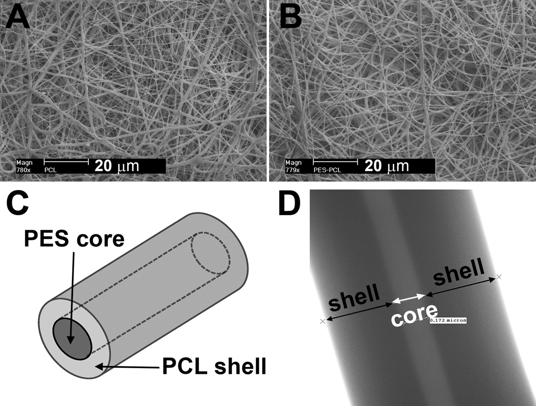
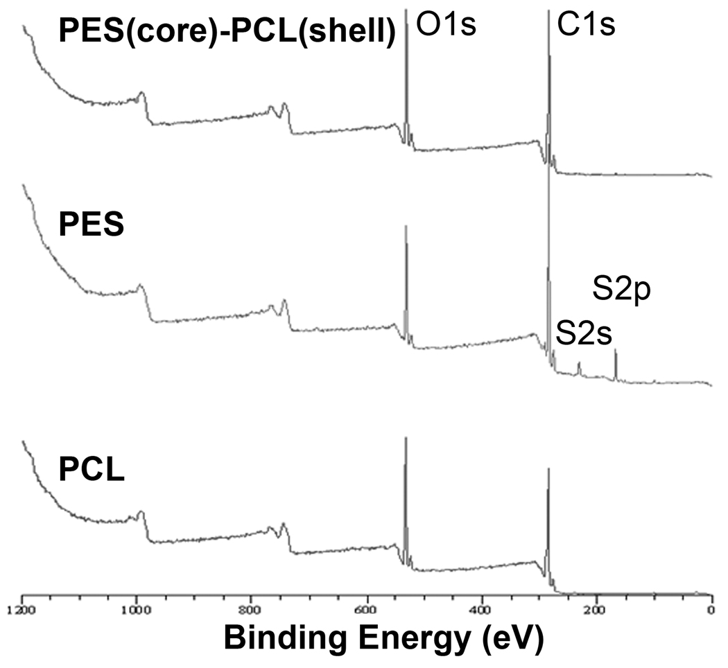
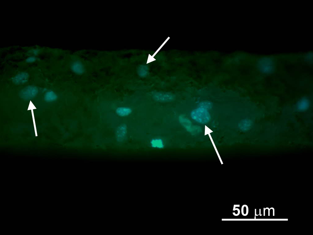
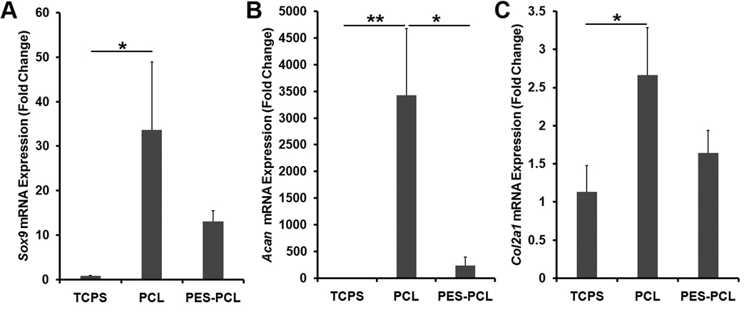
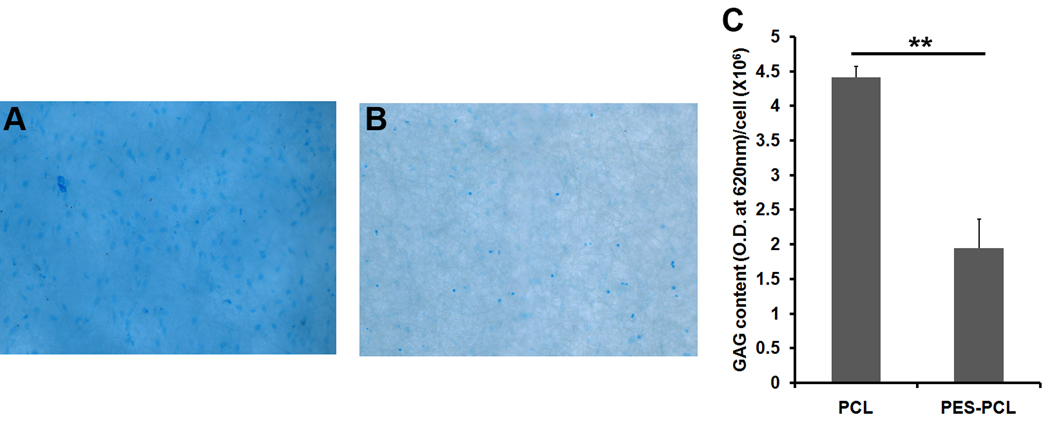
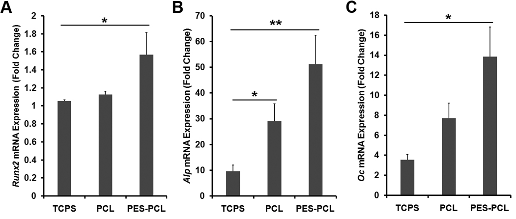
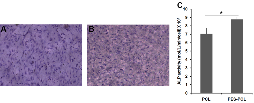
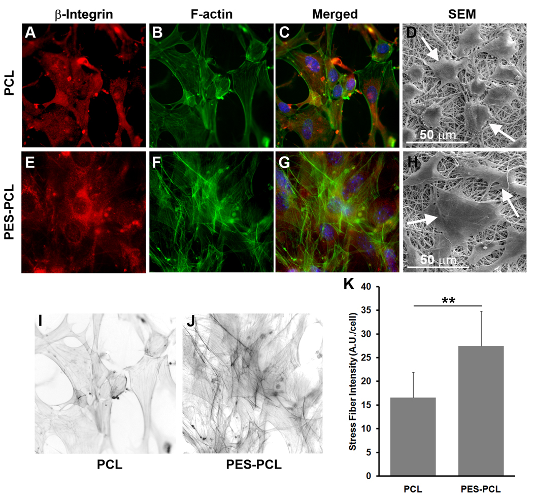
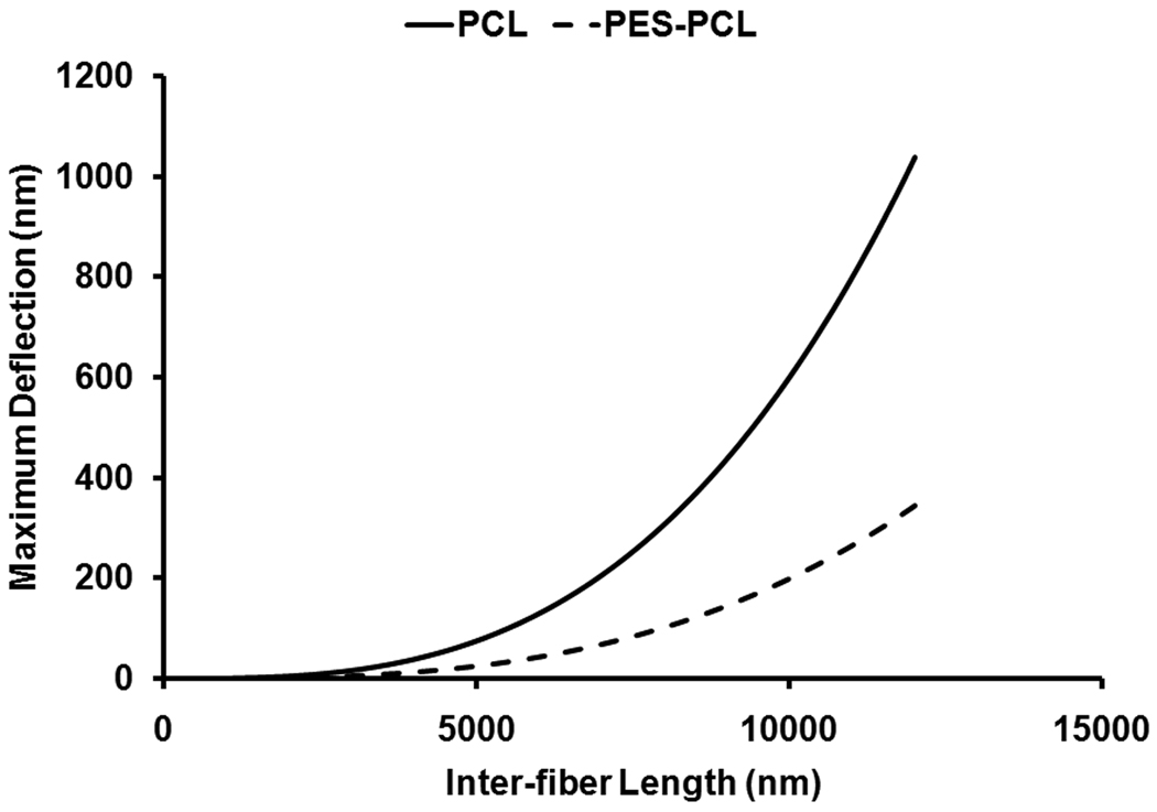
As the potential range of stem cell applications in tissue engineering continues to grow, the appropriate scaffolding choice is necessary to create tightly defined artificial microenvironments for each target organ. These microenvironments determine stem cell fate via control over differentiation. In this study we examined the specific effects of scaffold stiffness on embryonic mesenchymal progenitor cell behavior. Mechanically distinct scaffolds having identical microstructures and surface chemistries were produced utilizing core-shell electrospinning. The modulus of core-shell poly(ether sulfone)-poly(ε-caprolactone) (PES-PCL) fibers (30.6 MPa) was more than four times that of pure PCL (7.1 MPa). The results for chondrogenic and osteogenic differentiation of progenitor cells on each scaffold indicate that the lower modulus PCL fibers provided more appropriate microenvironments for chondrogenesis, evident by a marked up-regulation of chondrocytic Sox9, collagen type 2, and aggrecan gene expression and chondrocyte-specific extracellular matrix glycosaminoglycan production. In contrast, the stiffer core-shell PES-PCL fibers supported enhanced osteogenesis by promoting osteogenic Runx2, alkaline phosphatase, and osteocalcin gene expression, as well as alkaline phosphatase activity. The findings demonstrate that the microstructural stiffness/modules of a scaffold and the pliability of individual fibers may play a critical role in controlling stem cell differentiation. Regulation of cytoskeletal organization may occur via a "dynamic scaffold" leading to the subsequent intracellular signaling events that control differentiation-specific gene expression.


Figures

Figure 1
Figure 1
The microstructure of (A) pure PCL and (B) coaxial PES-PCL ‘core-shell’ electrospun fibers. (C) a schematic illustrates the microstructure of a core-shell fiber. (D) TEM image of a single ‘core-shell’ PES-PCL fiber showing a continuous PES core surrounded by a PCL shell.
Figure 2
Figure 2
XPS spectra of core-shell PES-PCL, pure PES and pure PCL fibers. O1s, C1s, S2s and S2p denote peaks detected from 1s atomic subshell of oxygen, 1s subshell of carbon, 2s subshell of sulfur and 2p subshell of sulfur, respectively.
Figure 3
Figure 3
A cross-sectional image of cell/scaffold constructs showing cellular infiltration into the scaffold. Arrows indicate nuclei (blue) in the electrospun matrix with green autofluorescence.
Figure 4
Figure 4
Chondrogenic ((A) Sox9, (B) aggrecan (Acan) and (C) collagen type 2 (Col2a)) gene expression of mesenchymal progenitor cells cultured on pure PCL and core-shell PES-PCL electrospun fibers under chondrogenic conditions as compared by real time-polymerase chain reaction (rt-PCR). Gene expression of the cells cultured on tissue culture polystyrene (TCPS) in chondrogenic conditions was used as a control (n=6; *: p < 0.05; **: p < 0.01). Expression level of each condition was normalized to the undifferentiated cells cultured on tissue culture plate.
Figure 5
Figure 5
Mesenchymal progenitor cells cultured on (A) pure PCL and (B) core-shell PES-PCL electrospun fibers under chondrogenic conditions stained with Alcian blue showing greater glycosaminoglycan (GAG) production on PCL fibers in comparison to core-shell fibers. Images were taken at 100X. (C) Spectrophotometric analysis of GAG synthesis by the cells grown on pure PCL fibers and core-shell PES-PCL electrospun fibers. (n=6; **: p < 0.01).
Figure 6
Figure 6
Osteogenic ((A) Runx2, (B) alkaline phosphatase (Alp), and (C) osteocalcin (Oc)) gene expression of mesenchymal progenitor cells cultured on pure PCL and core-shell PES-PCL electrospun fibers under osteogenic conditions as compared by real time-polymerase chain reaction (rt-PCR). Gene expression of the cells cultured on tissue culture polystyrene (TCPS) in osteogenic conditions was used as a control (n=6, *: p < 0.05; **: p < 0.01). Expression level of each condition was normalized to the undifferentiated cells cultured on tissue culture plate.
Figure 7
Figure 7
Mesenchymal progenitor cells cultured on (A) pure PCL and (B) core-shell PES-PCL electrospun fibers under osteogenic conditions and stained to assess alkaline phosphatase activity (enzyme: pink to red, nuclei: blue). Images were taken at 100X. (C) Quantitative analysis of alkaline phosphatase activity by spectrophotometry in the cells grown on the pure PCL and core-shell fibers (n=6; *: p < 0.05, **: p < 0.01).
Figure 8
Figure 8
The intracellular structure of mesenchymal progenitor cells on pure PCL (A–D) and core-shell PES-PCL (E–H) electrospun fibers under chondrogenic conditions by immunofluorescent analysis of the adhesion molecule integrin β1 (red, A and E), F-actin (green, B and F), and SEM (D and H). The cells on both substrates show similarly uniform intracellular distribution of integrin β1. However, the stiffness of the fibers significantly affected stress fiber organization. SEM examination shows more rounded cellular morphology on pure PCL (D) compared to the spread morphology on PES-PCL (H) fibers. (I, J) Inverted images of F-actin staining exhibiting greater development of stress fibers in the cells grown on the core-shell (J) versus pure PCL (I) scaffolds. (K) Quantitative analysis of the F-actin stained images to derive the mean intensity of stress fibers per cell basis (A.U.: arbitrary unit; n = 18; **: p < 0.01). Images were taken at 400X.
Figure 9
Figure 9
Theoretical deflection (under 75 nN of force) of an individual electrospun PCL (solid line) or PES-PCL (dashed line) fiber versus inter-fiber distance (Table 2). The pure PCL fiber can be moved significantly greater distances by adherent cells relative to the core-shell PES-PCL fiber.

Similar articles

Cited by

References

    1. Park IH, Zhao R, West JA, Yabuuchi A, Huo HG, Ince TA, et al. Reprogramming of human somatic cells to pluripotency with defined factors. Nature. 2008;451 141-U1. - PubMed
    1. Takahashi K, Tanabe K, Ohnuki M, Narita M, Ichisaka T, Tomoda K, et al. Induction of pluripotent stem cells from adult human fibroblasts by defined factors. Cell. 2007;131:861–872. - PubMed
    1. Bianco P, Riminucci N, Kuznetsov S, Robey PG. Multipotential cells in the bone marrow stroma: Regulation in the context of organ physiology. Critical Reviews in Eukaryotic Gene Expression. 1999;9:159–173. - PubMed
    1. Prockop DJ. Marrow stromal cells as steam cells for nonhematopoietic tissues. Science. 1997;276:71–74. - PubMed
    1. Sun N, Panetta NJ, Gupta DM, Wilson KD, Lee A, Jia F, et al. Feeder-free derivation of induced pluripotent stem cells from adult human adipose stem cells. Proceedings of the National Academy of Sciences of the United States of America. 2009;106:15720–15725. - PMC - PubMed

Publication types

MeSH terms