. 2010 Dec 10;87(6):882-9.
doi: 10.1016/j.ajhg.2010.10.026. Epub 2010 Nov 25.

A homozygous mutation in the tight-junction protein JAM3 causes hemorrhagic destruction of the brain, subependymal calcification, and congenital cataracts

Affiliations

A homozygous mutation in the tight-junction protein JAM3 causes hemorrhagic destruction of the brain, subependymal calcification, and congenital cataracts

Ganeshwaran H Mochida et al. Am J Hum Genet. .

Abstract

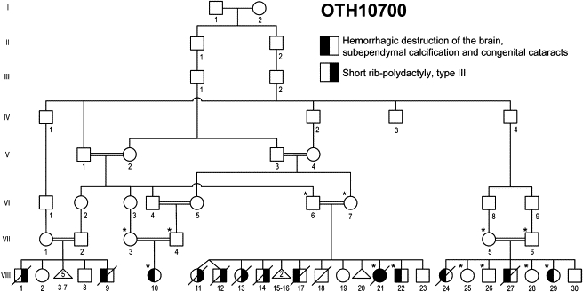
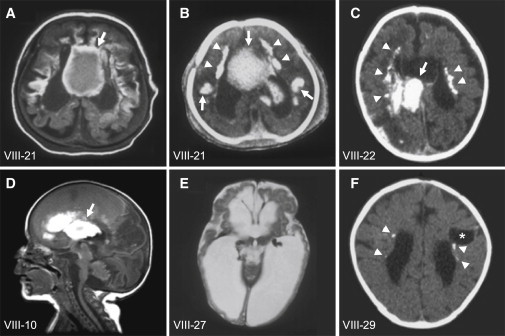
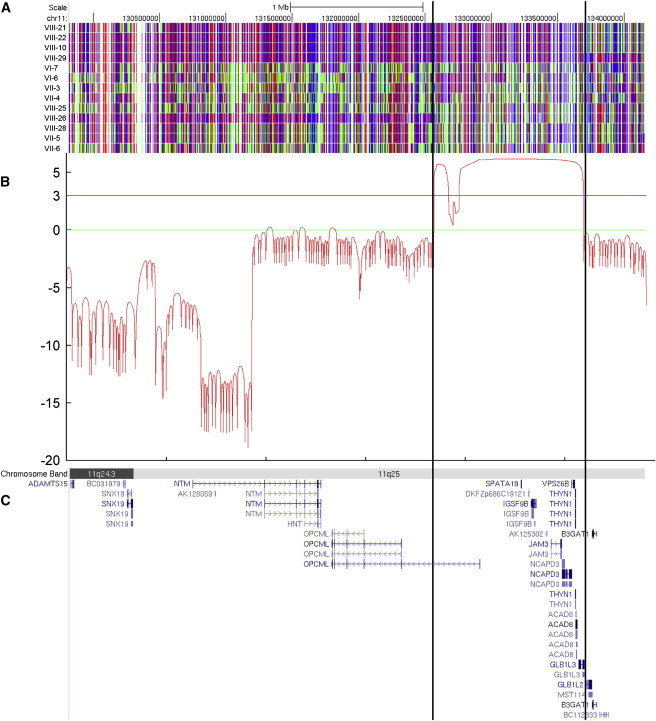
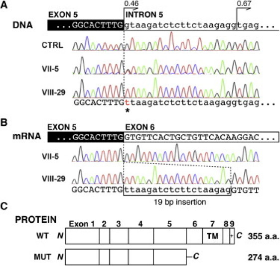
The tight junction, or zonula occludens, is a specialized cell-cell junction that regulates epithelial and endothelial permeability, and it is an essential component of the blood-brain barrier in the cerebrovascular endothelium. In addition to functioning as a diffusion barrier, tight junctions are also involved in signal transduction. In this study, we identified a homozygous mutation in the tight-junction protein gene JAM3 in a large consanguineous family from the United Arab Emirates. Some members of this family had a rare autosomal-recessive syndrome characterized by severe hemorrhagic destruction of the brain, subependymal calcification, and congenital cataracts. Their clinical presentation overlaps with some reported cases of pseudo-TORCH syndrome as well as with cases involving mutations in occludin, another component of the tight-junction complex. However, massive intracranial hemorrhage distinguishes these patients from others. Homozygosity mapping identified the disease locus in this family on chromosome 11q25 with a maximum multipoint LOD score of 6.15. Sequence analysis of genes in the candidate interval uncovered a mutation in the canonical splice-donor site of intron 5 of JAM3. RT-PCR analysis of a patient lymphoblast cell line confirmed abnormal splicing, leading to a frameshift mutation with early termination. JAM3 is known to be present in vascular endothelium, although its roles in cerebral vasculature have not been implicated. Our results suggest that JAM3 is essential for maintaining the integrity of the cerebrovascular endothelium as well as for normal lens development in humans.


Figures

Figure 1
Figure 1
Pedigree Drawing of the Family OTH10700 Filled symbols denote affected individuals. The left half of the symbol is filled for the syndrome of hemorrhagic destruction of the brain, subependymal calcification, and congenital cataracts, and the right half of the symbol is filled for short rib-polydactyly syndrome, type III. Genotyped individuals are indicated with an asterisk.
Figure 2
Figure 2
Brain Imaging of the Family OTH10700 A T1-weighted axial brain MRI (A) and brain CT (B) of individual VIII-21, a brain CT of individual VIII-22 (C), and a T1-weighted sagittal brain MRI of individual VIII-10 (D) show multifocal intraparenchymal hemorrhage, predominantly in the cerebral white matter and basal ganglia (arrows). A T2-weighted axial brain MRI of individual VIII-27 (E) shows massive cystic destruction of the cerebral white matter and basal ganglia and resulting large lateral ventricles. A brain CT of individual VIII-29 (F) shows a porencephalic cyst centered in the left frontal subcortical white matter (asterisk), enlarged lateral ventricles, and reduced white-matter volume, indicative of destructive changes. Subependymal calcification is evident on CT (B, C and F, arrowheads). Figure 2A is from the same MRI series previously published in Al-Gazali et al. (this individual is referred to as case 4 in the paper).
Figure 3
Figure 3
Homozygosity Mapping and Linkage Analysis of the Family OTH10700 (A) SNP genotyping results on chromosome 11q25. A block of homozygosity shared only by the affected individuals tested (VIII-21, VIII-22, VIII-10, and VIII-29) is evident and delineated by the vertical lines. Red and blue indicate homozygous SNP markers, and green indicates heterozygous SNP markers. (B) LOD score graph of the candidate region. A maximum multipoint LOD score of 6.15 was obtained within the candidate interval. (C) Genes within the candidate region, shown as the RefSeq track of the UCSC Genome Browser (NCBI build 36/hg18).
Figure 4
Figure 4
Mutation in JAM3 (A) Genomic DNA analysis of JAM3. Affected individual VIII-29 shows a G-to-T change in the canonical splice donor site of JAM3 intron 5 (c.747+1G>T; asterisk). Her mother (VII-5) is heterozygous for the mutation. A cryptic splice donor site 19 bp downstream is at least equally as potent as the wild-type donor site (confidence score 0.46 [wild-type] versus 0.67 [cryptic] by NetGene2). CTRL = normal control. (B) mRNA analysis of JAM3. On the mRNA level, the mutation results in the usage of the downstream cryptic splice donor site and causes a 19 bp insertion in the affected individual (VIII-29). Only normal splice products are seen in her mother (VII-5). (C) Predicted protein product of the mutant allele. The 19 bp insertion is expected to result in a truncating protein product with 25 aberrant amino acid residues in the C terminus (WT = wild-type protein, MUT = mutant protein). The truncation occurs proximal to the transmembrane domain (TM) and PDZ-binding motif (+).

References

    1. Ebnet K. Organization of multiprotein complexes at cell-cell junctions. Histochem. Cell Biol. 2008;130:1–20. - PMC - PubMed
    1. Vestweber D. Molecular mechanisms that control endothelial cell contacts. J. Pathol. 2000;190:281–291. - PubMed
    1. Carlton V.E., Harris B.Z., Puffenberger E.G., Batta A.K., Knisely A.S., Robinson D.L., Strauss K.A., Shneider B.L., Lim W.A., Salen G. Complex inheritance of familial hypercholanemia with associated mutations in TJP2 and BAAT. Nat. Genet. 2003;34:91–96. - PubMed
    1. Wilcox E.R., Burton Q.L., Naz S., Riazuddin S., Smith T.N., Ploplis B., Belyantseva I., Ben-Yosef T., Liburd N.A., Morell R.J. Mutations in the gene encoding tight junction claudin-14 cause autosomal recessive deafness DFNB29. Cell. 2001;104:165–172. - PubMed
    1. Riazuddin S., Ahmed Z.M., Fanning A.S., Lagziel A., Kitajiri S., Ramzan K., Khan S.N., Chattaraj P., Friedman P.L., Anderson J.M. Tricellulin is a tight-junction protein necessary for hearing. Am. J. Hum. Genet. 2006;79:1040–1051. - PMC - PubMed

Publication types

Substances