

Dominant mutations in KBTBD13, a member of the BTB/Kelch family, cause nemaline myopathy with cores
- PMID: 21109227
- PMCID: PMC2997379
- DOI: 10.1016/j.ajhg.2010.10.020
Dominant mutations in KBTBD13, a member of the BTB/Kelch family, cause nemaline myopathy with cores
Erratum in
- Am J Hum Genet. 2011 Jan 7;88(1):122
Abstract
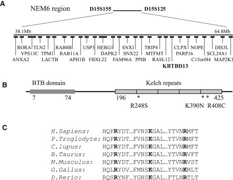
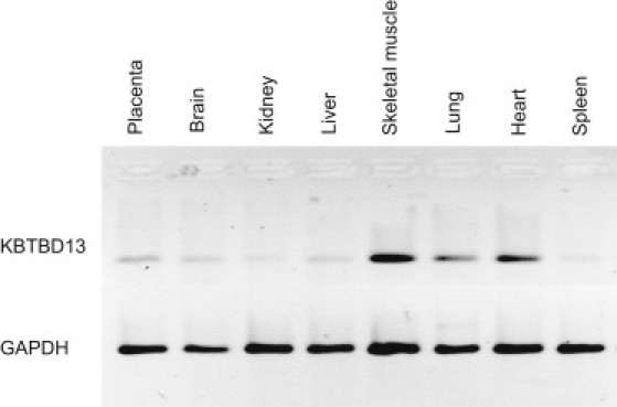
We identified a member of the BTB/Kelch protein family that is mutated in nemaline myopathy type 6 (NEM6), an autosomal-dominant neuromuscular disorder characterized by the presence of nemaline rods and core lesions in the skeletal myofibers. Analysis of affected families allowed narrowing of the candidate region on chromosome 15q22.31, and mutation screening led to the identification of a previously uncharacterized gene, KBTBD13, coding for a hypothetical protein and containing missense mutations that perfectly cosegregate with nemaline myopathy in the studied families. KBTBD13 contains a BTB/POZ domain and five Kelch repeats and is expressed primarily in skeletal and cardiac muscle. The identified disease-associated mutations, C.742C>A (p.Arg248Ser), c.1170G>C (p.Lys390Asn), and c.1222C>T (p.Arg408Cys), located in conserved domains of Kelch repeats, are predicted to disrupt the molecule's beta-propeller blades. Previously identified BTB/POZ/Kelch-domain-containing proteins have been implicated in a broad variety of biological processes, including cytoskeleton modulation, regulation of gene transcription, ubiquitination, and myofibril assembly. The functional role of KBTBD13 in skeletal muscle and the pathogenesis of NEM6 are subjects for further studies.
Copyright © 2010 The American Society of Human Genetics. Published by Elsevier Inc. All rights reserved.
Figures

References
-
- Ryan M.M., Schnell C., Strickland C.D., Shield L.K., Morgan G., Iannaccone S.T., Laing N.G., Beggs A.H., North K.N. Nemaline myopathy: a clinical study of 143 cases. Ann. Neurol. 2001;50:312–320. - PubMed
-
- Sanoudou D., Beggs A.H. Clinical and genetic heterogeneity in nemaline myopathy—a disease of skeletal muscle thin filaments. Trends Mol. Med. 2001;7:362–368. - PubMed
-
- Laing N.G., Wallgren-Pettersson C. 161st ENMC International Workshop on nemaline myopathy and related disorders, Newcastle upon Tyne, 2008. Neuromuscul Disord. 2009;19:300–305. - PubMed
-
- North K. What's new in congenital myopathies? Neuromuscul. Disord. 2008;18:433–442. - PubMed
Publication types
MeSH terms
Substances
Grants and funding
LinkOut - more resources
Full Text Sources
Molecular Biology Databases
