. 2011 Apr;39(7):2799-808.
doi: 10.1093/nar/gkq1220. Epub 2010 Nov 24.

Endogenous ribosomal frameshift signals operate as mRNA destabilizing elements through at least two molecular pathways in yeast

Affiliations

Endogenous ribosomal frameshift signals operate as mRNA destabilizing elements through at least two molecular pathways in yeast

Ashton T Belew et al. Nucleic Acids Res. 2011 Apr.

Abstract

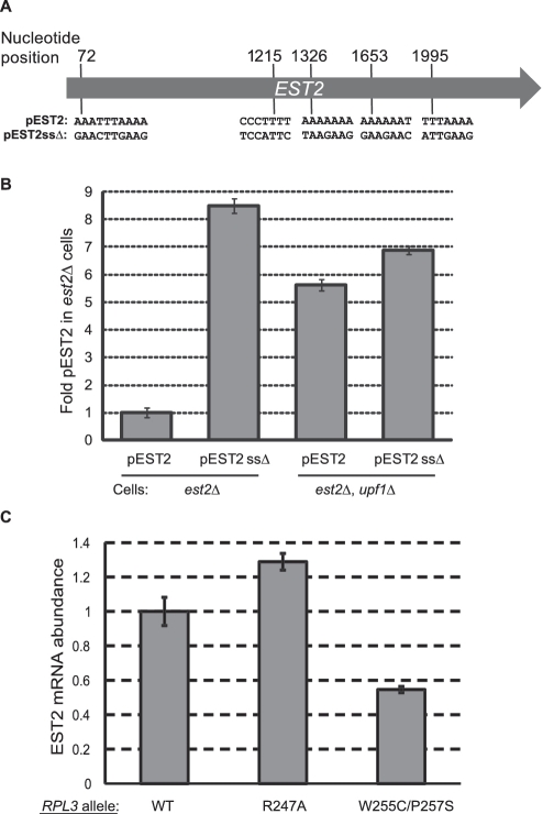
Although first discovered in viruses, previous studies have identified operational -1 ribosomal frameshifting (-1 RF) signals in eukaryotic genomic sequences, and suggested a role in mRNA stability. Here, four yeast -1 RF signals are shown to promote significant mRNA destabilization through the nonsense mediated mRNA decay pathway (NMD), and genetic evidence is presented suggesting that they may also operate through the no-go decay pathway (NGD) as well. Yeast EST2 mRNA is highly unstable and contains up to five -1 RF signals. Ablation of the -1 RF signals or of NMD stabilizes this mRNA, and changes in -1 RF efficiency have opposing effects on the steady-state abundance of the EST2 mRNA. These results demonstrate that endogenous -1 RF signals function as mRNA destabilizing elements through at least two molecular pathways in yeast. Consistent with current evolutionary theory, phylogenetic analyses suggest that -1 RF signals are rapidly evolving cis-acting regulatory elements. Identification of high confidence -1 RF signals in ∼10% of genes in all eukaryotic genomes surveyed suggests that -1 RF is a broadly used post-transcriptional regulator of gene expression.


Figures

Figure 1.
Figure 1.
Schematic of PGK1 reporter vectors used to monitor the effects of −1 RF signals on mRNA stability. The indicated Renilla and firefly luciferase derived sequences from pJD375 were cloned into the unique KpnI restriction site in a high copy PGK1 expression vector to create the readthrough control (pJD753). The indicated −1 RF signals derived from BUB3, EST2, SPR6 and TBF1 were cloned into SalI/BamHI digested pJD753. Colored arcs depict computationally predicted base-paired stems (17). The premature termination control (PTC) was constructed by mutagenizing pJD753 to create an in-frame TAA codon.
Figure 2.
Figure 2.
Cellular −1 RF signals can decrease mRNA steady-state abundance in yeast. The −1 RF signals from SPR6, EST2, BUB3 and TBF1 were cloned into a PGK1 reporter minigene such that frameshift events would cause elongating ribosomes to encounter premature termination codons (PTC). Readthrough (RT) and in-frame PTC containing reporters are included as controls. Northern blots of total mRNAs extracted from logarithmically growing cells were probed with a reporter-specific oligonucleotide (PGK), stripped and re-hybridized with a U3 snoRNA-specific probe for normalization. All blots were repeated at least three times. (A) Steady-state abundance of reporter mRNAs in wild-type cells. Graph shows abundances of test mRNAs relative to the readthrough control. (B) Same as panel A, but in upf1Δ cells. Graph plots abundance of specific test mRNAs in upf1Δ versus wild-type cells. (C–F) These panels are similar to panel B, except that samples were extracted from dom34Δ, dcp1Δ, xrn1Δ and ski3Δ cells respectively. Bars denote standard errors.
Figure 3.
Figure 3.
The EST2 −1 RF signal at position 1652 destabilizes mRNA primarily through NMD. (A–H) The readthrough control, in-frame PTC control and EST2 −1 RF containing PGK1 reporters were introduced into either wild-type (A–D) or upf1Δ (E–H) cells harboring the temperature-sensitive rpb1-1 allele of RNA polymerase II. Total mRNAs were harvested from cells after temperature shift at the indicated timepoints, and Northern blots were probed using the PGK1 reporter-specific and U3 snoRNA specific probes. Graph in panel D plots normalized PGK1 reporter mRNA abundances in wild-type cells, and graph in panel h plots these data in upf1Δ cells. (I) The wild-type A AAA AAT slippery site of the EST2 −1 RF signal in the PGK1 reporter was changed to G AAG AAC, and steady state northern blot analyses were performed using mRNAs extracted from cells expressing the readthrough control, the in-frame PTC containing control, and cells expressing either the wild-type or mutant slippery sites. WTss denotes the wild-type slippery site. SSm denotes the slippery site mutant. DOM34 and dom34Δ denote isogenic wild-type and dom34Δ cells. Fold-RT denotes fold readthrough control. SD (±) denotes standard deviation.
Figure 4.
Figure 4.
Silent coding mutations that disrupt 5 slippery sites in the EST2 gene stabilize its mRNA. (A) Schematic of the EST2 coding sequence. Positions of the slippery sites of five predicted −1 RF signals and their sequences are indicated. The full-length gene including native 5′ and 3′ UTR sequences were cloned into a low-copy yeast vector to create pEST2. Silent coding mutations that are predicted to inactivate −1 RF were introduced to produce pEST2ssΔ. (B) pEST2 or pEST2ssΔ were introduced into est2Δ or est2Δ upf1Δ cells and EST2 mRNA steady state abundances were determined by quantitative real-time PCR. (C) Quantitative real-time PCR was used to monitor steady-state abundance of the endogenous EST2 mRNA in isogenic cells expressing three different forms of ribosomal protein L3: Wild-Type RPL3 (WT); the R247A mutant which promotes decreased rates of −1 RF and the W255C/P257S mutant which promotes increased −1 RF efficiency. EST2 mRNA abundances were normalized to U3 snoRNA abundance for each sample, and the values shown are relative to wild-type cells.
Figure 5.
Figure 5.
−1 RF signals can function as mRNA destabilizing elements through the nonsense-mediated mRNA and NGD Pathways. Left panel (A) −1 RF event directs an elongating ribosome to encounter a premature termination codon (PTC). This leads to recruitment of the surveillance complex (Upf proteins), leading to mRNA decapping and 5′ → 3′ degradation by Xrn1p and deadenylation and 3′ → 5′ degradation by the degradasome. Right panel: the mRNA pseudoknot in a −1 RF signal causes elongating ribosomes to pause, recruiting the Dom34p/Hbs1p complex, thus initiating NGD.

References

    1. Brierley I. Ribosomal frameshifting on viral RNAs. J. Gen. Virol. 1995;76:1885–1892. - PubMed
    1. Farabaugh PJ. Programmed translational frameshifting. Microbiol. Rev. 1996;60:103–134. - PMC - PubMed
    1. Baranov PV, Gesteland RF, Atkins JF. Recoding: translational bifurcations in gene expression. Gene. 2002;286:187–201. - PubMed
    1. Kontos H, Napthine S, Brierley I. Ribosomal pausing at a frameshifter RNA pseudoknot is sensitive to reading phase but shows little correlation with frameshift efficiency. Mol. Cell. Biol. 2001;21:8657–8670. - PMC - PubMed
    1. Lopinski JD, Dinman JD, Bruenn JA. Kinetics of ribosomal pausing during programmed -1 translational frameshifting. Mol. Cell. Biol. 2000;20:1095–1103. - PMC - PubMed

Publication types

MeSH terms