. 2011 Jan;12(1):63-70.
doi: 10.1038/embor.2010.170. Epub 2010 Nov 26.

The hypoxia-inducible transcription factor pathway regulates oxygen sensing in the simplest animal, Trichoplax adhaerens

Affiliations

The hypoxia-inducible transcription factor pathway regulates oxygen sensing in the simplest animal, Trichoplax adhaerens

Christoph Loenarz et al. EMBO Rep. 2011 Jan.

Abstract

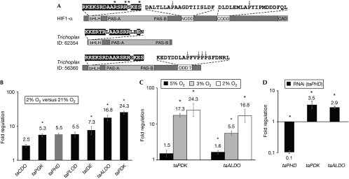
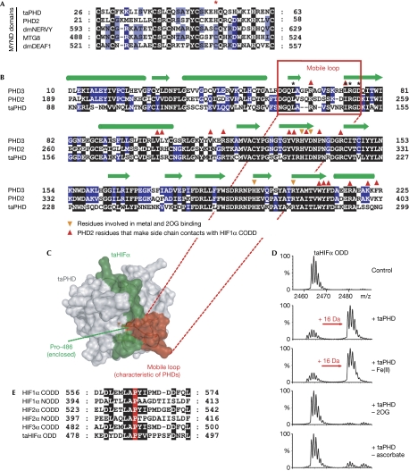
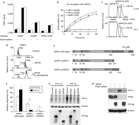
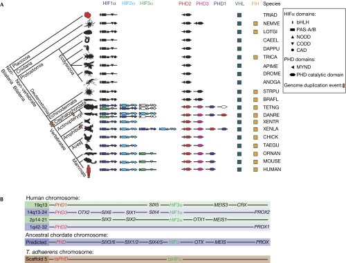
The hypoxic response in humans is mediated by the hypoxia-inducible transcription factor (HIF), for which prolyl hydroxylases (PHDs) act as oxygen-sensing components. The evolutionary origins of the HIF system have been previously unclear. We demonstrate a functional HIF system in the simplest animal, Trichoplax adhaerens: HIF targets in T. adhaerens include glycolytic and metabolic enzymes, suggesting a role for HIF in the adaptation of basal multicellular animals to fluctuating oxygen levels. Characterization of the T. adhaerens PHDs and cross-species complementation assays reveal a conserved oxygen-sensing mechanism. Cross-genomic analyses rationalize the relative importance of HIF system components, and imply that the HIF system is likely to be present in all animals, but is unique to this kingdom.


Conflict of interest statement

The authors declare that they have no conflict of interest.

Figures

Figure 1
Figure 1
Hypoxic regulation of Trichoplax adhaerens genes. (A) Domain structures of T. adhaerens bHLH-PAS proteins; sequence 56360 (Srivastava et al, 2008) corresponds to the likely taHIFα homologue. Asterisks indicate (predicted) DNA-interacting residues on the basis of homology modelling (PDB 1AN4). Known and putative HIFα ODD sequences are shown; arrows indicate known and putative prolyl-hydroxylation sites. (B) RT–qPCR analysis of T. adhaerens showing fold regulation in hypoxia (2% O2) relative to normoxia (normalized to β-actin; n=3; ±s.e.m.; *P<0.05). (C) Increasing degrees of hypoxia increased expression of taALDO and taPDK (normalized to β-actin; n=3; ±s.e.m.; *P<0.05). (D) RT–qPCR analysis of RNAi against taPHD in T. adhaerens. Whereas taPHD levels are reduced, expression of hypoxia-inducible genes (taPDK and taALDO) is increased (normalized to β-actin; n=3; ±s.e.m.; *P<0.05). ALDO, fructose-biphosphate aldolase; bHLH, basic helix–loop–helix; CDO, cysteine dioxygenase; HIF1α, hypoxia-inducible transcription factor 1α; IDE, insulin-degrading enzyme; ODD, oxygen-dependent degradation domain; PDK, pyruvate dehydrogenase kinase; PGK, phosphoglycerate kinase; PLOD, procollagen lysine hydroxylase; RNAi, RNA interference; RT–qPCR, reverse transcription–quantitative PCR; taHIFα, Trichoplax adhaerens hypoxia-inducible transcription factor-α; taPHD, Trichoplax adhaerens prolyl-hydroxylase.
Figure 2
Figure 2
Trichoplax adhaerens PHD has conserved substrate-binding features and is active as a taHIFα prolyl hydroxylase. (A) Comparison of the MYND finger for PHD2/taPHD with stereotypical MYND finger sequences. Note that one of the cysteines is replaced by a histidine in the PHDs (indicated by an asterisk; GenBank entries: dmNERVY, 45445680; MTG8, 4757916; dmDEAF1, 7293736). (B) Sequence comparison of human PHD2/3 with taPHD; sequences corresponding to the mobile region are boxed. Secondary structures: α-helices, green cylinders; β-strands, green arrows. (C) Homology model for the binding of taHIFα (green) by taPHD (grey; using PDB 3HQR). Note that a mobile loop (red) is conserved in taPHD and appears ‘anchored' to the active site by electrostatic (Arg 148/Asp 150) and hydrophobic interactions (Leu 138/Ile 147). (D) Mass spectrometric analyses showing taPHD-catalysed hydroxylation of taHIFα ODD (16-Da mass shift). taPHD co-purifies with Fe(II) and its activity is stimulated by ascorbate. (E) Alignment of T. adhaerens and human HIFα ODD domains. CODD, C-terminal ODD; NODD, N-terminal ODD; ODD, oxygen-dependent degradation domain; 2OG, 2-oxoglutarate; PHD, prolyl hydroxylase; taHIFα, T. adhaerens hypoxia-inducible transcription factor-α; taPHD, T. adhaerens prolyl-hydroxylase.
Figure 3
Figure 3
T. adhaerens prolyl-hydroxylase and prolyl hydroxylase 2 have conserved functions in the hypoxic response. (A) FRET assay showing that binding of human and T. adhaerens HIFα peptides to the VHL complex depends on prolyl trans-4-hydroxylation (two replicates). (B) taPHD-catalysed hydroxylation of an equimolar mixture of HIF1α NODD, CODD and taHIFα ODD (NODD hydroxylation was observed after incubation overnight). (C) Incubation of taPHD with equimolar Fe(II) and 2OG (mass: 146 Da) leads to a stable complex in the absence of substrate (half-life >24 h; note the lack of succinate formation), as shown by non-denaturing electrospray ionization mass spectrometry. (D) Hydroxylation of taHIFα in 18O2 proceeds with 18O incorporation. (E) Domain analysis of taHIFα splice variants. (F) Reverse transcription–quantitative PCR analysis of the effect of hypoxia on relative taHIFα splice variant levels (n=3; ±s.e.m.; *P<0.05). See supplementary Fig S4 online for splice sites. (G) Immunoblot of human 293T cells transfected with haemagglutinin-tagged human PHD22−426, C. elegans EGL92−723 and T. adhaerens taPHD, showing that all enzymes cause reduction in endogenous HIF1α levels in hypoxia. (H) Immunoblot of human 293T cells showing that taPHD is sufficient to suppress endogenous HIF1α levels in the absence of PHD2. Cells were transfected with or without haemagglutinin-tagged T. adhaerens taPHD plus control or PHD2 siRNA. CODD, C-terminal ODD; FRET, fluorescence resonance energy transfer; HA, haemagglutinin; HIFα, hypoxia-inducible transcription factor-α; NODD, N-terminal oxygen-dependent degradation domain; 2OG, 2-oxoglutarate; PHD, prolyl hydroxylase; siRNA, small interfering RNA; taPHD, T. adhaerens prolyl-hydroxylase; VHL, von Hippel Lindau protein.
Figure 4
Figure 4
Evolutionary analysis of the hypoxia-inducible transcription factor system. (A) Phylogenetic domain analysis of HIF system gene products across metazoans (not to scale). Ovals represent genome duplications; likely pseudogenes are not shown; uncoloured domains indicate no isoform assignment; grey domains reflect predictions necessitated by incompletely sequenced genomes. Profile HMMs were used to search for more distant homologues. In some deposited genomes, protein domains of interest were annotated as introns (possibly due, in part, to their low sequence conservation), requiring both gene re-annotation and profile HMM searches considering all likely genome translations. See supplementary Tables S2 and S3 online for sequences of HIFα subdomains and abbreviations. (B) Comparison of the relative locations of human HIFα and PHD genes (not to scale), with prediction of their relative position in an ancestral chordate genome, and T. adhaerens gene location (note that basal animals contain few homeobox genes). In the human genome, HIFα and PHD genes map to four related chromosome regions, close to homeobox genes of the SIX1/2, SIX3/6, SIX4/5, PROX, MEIS and OTX gene families. bHLH, basic helix–loop–helix; CAD, C-terminal transcriptional activation domain; CODD, C-terminal oxygen-dependent degradation domain; FIH, factor inhibiting HIF; HIF, hypoxia-inducible transcription factor; HMM, hidden Markov model; NODD, N-terminal ODD; PAS, Per–Arnt–Sim; PHD, prolyl hydroxylase; VHL, von Hippel Lindau protein.

Comment in

References

    1. Abedin M, King N (2008) The premetazoan ancestry of cadherins. Science 319: 946–948 - PubMed
    1. Chan DA, Sutphin PD, Yen SE, Giaccia AJ (2005) Coordinate regulation of the oxygen-dependent degradation domains of hypoxia-inducible factor 1α. Mol Cell Biol 25: 6415–6426 - PMC - PubMed
    1. Chowdhury R, McDonough MA, Mecinovi J, Loenarz C, Flashman E, Hewitson KS, Domene C, Schofield CJ (2009) Structural basis for binding of hypoxia-inducible factor to the oxygen-sensing prolyl hydroxylases. Structure 17: 981–989 - PubMed
    1. Cioffi CL, Qin Liu X, Kosinski PA, Garay M, Bowen BR (2003) Differential regulation of HIF-1α prolyl-4-hydroxylase genes by hypoxia in human cardiovascular cells. Biochem Biophys Res Commun 303: 947–953 - PubMed
    1. Flashman E, Bagg EAL, Chowdhury R, Mecinovic J, Loenarz C, McDonough MA, Hewitson KS, Schofield CJ (2008) Kinetic rationale for selectivity toward N- and C-terminal oxygen-dependent degradation domain substrates mediated by a loop region of hypoxia-inducible factor prolyl hydroxylases. J Biol Chem 283: 3808–3815 - PubMed

Publication types

MeSH terms

Substances

LinkOut - more resources