. 2011 Jan;38(1):105-12.

Hsp90 inhibition increases p53 expression and destabilizes MYCN and MYC in neuroblastoma

Affiliations

Hsp90 inhibition increases p53 expression and destabilizes MYCN and MYC in neuroblastoma

Paul L Regan et al. Int J Oncol. 2011 Jan.

Abstract

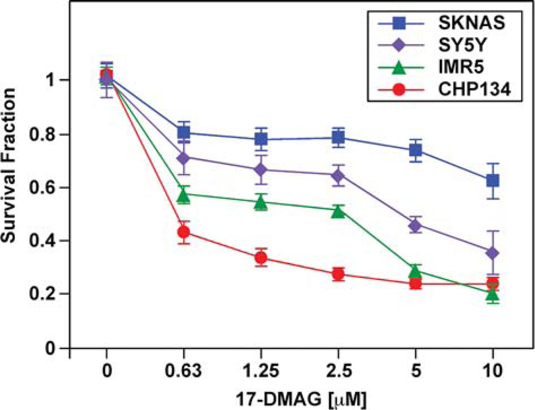
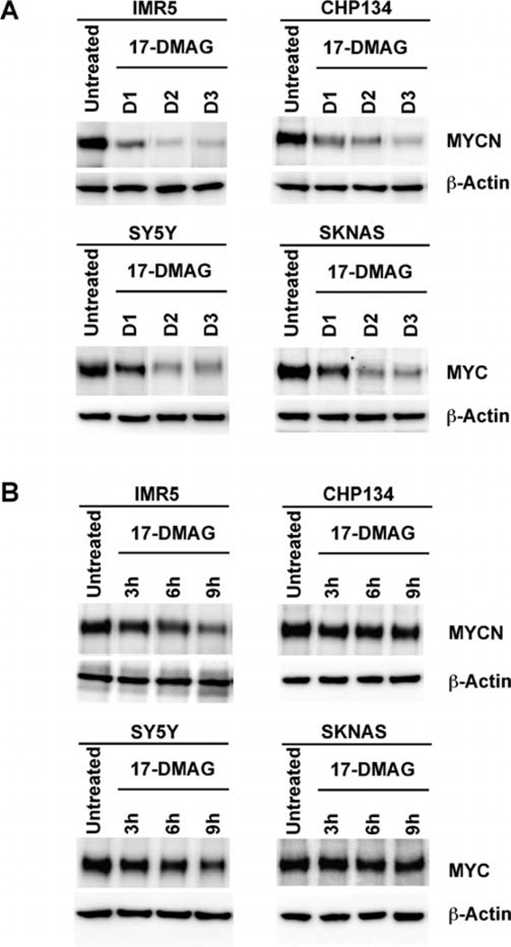
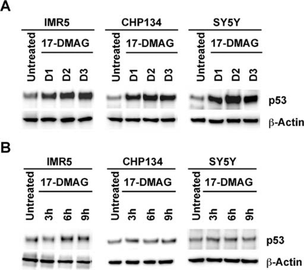
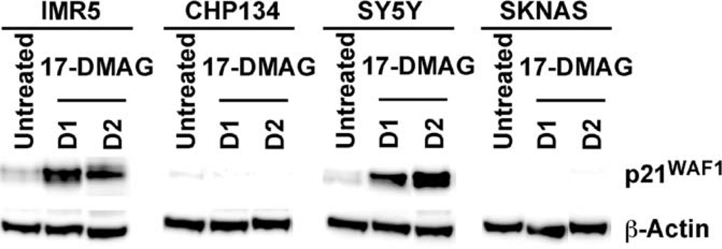
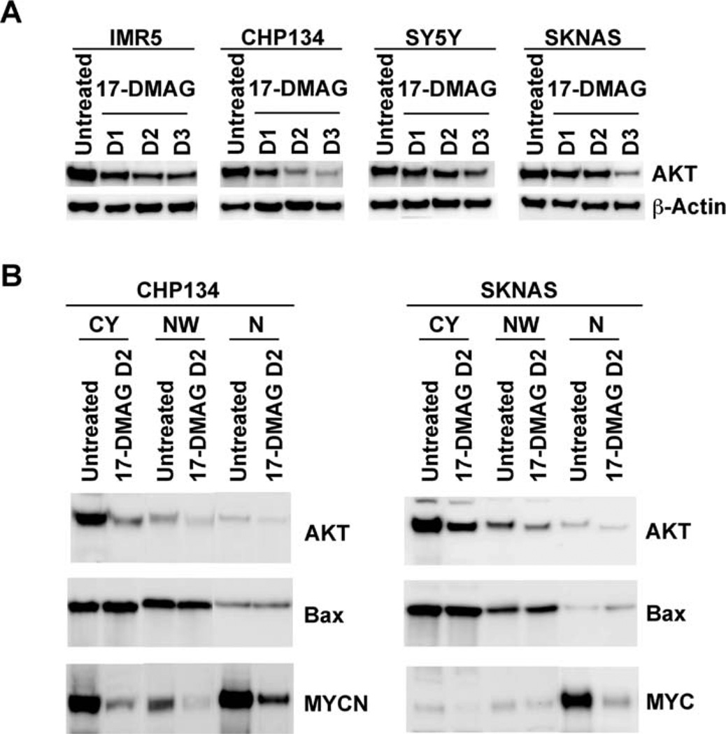
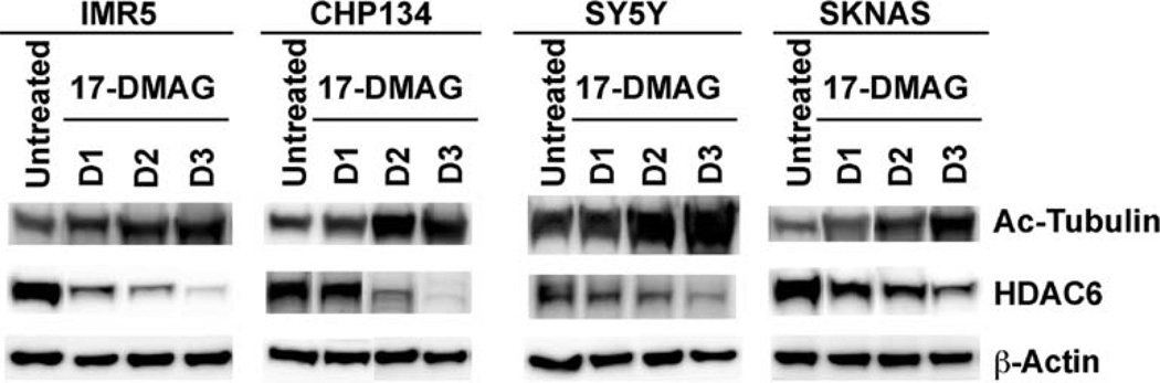
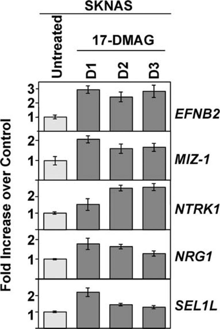
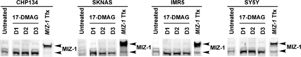
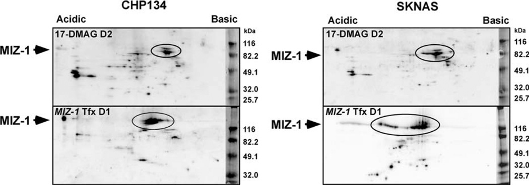
Neuroblastoma is a childhood cancer that exhibits either a favorable or an unfavorable phenotype. MYCN and MYC are oncoproteins that play crucial roles in determining the malignancy of unfavorable neuroblastoma. The Hsp90 superchaperone complex assists in the folding and function of a variety of oncogenic client proteins. Inhibition of Hsp90 by small molecule inhibitors leads to the destabilization of these oncogenic proteins and consequently suppresses tumor malignancy. Nonetheless, little is known about the effect of Hsp90 inhibition on the stability of MYCN and MYC proteins. In this study, we investigated the effect of Hsp90 inhibition on the phenotype of unfavorable neuroblastoma cells including its effect on MYCN and MYC expression. Two MYCN-amplified neuroblastoma cell lines (IMR5 and CHP134) and two non-MYCN-amplified cell lines (SY5Y and SKNAS) were used to address the effect of Hsp90 inhibition on the malignant phenotype of neuroblastoma. It was found that Hsp90 inhibition in neuroblastoma cell lines resulted in significant growth suppression, a decrease in MYCN and MYC expression, and an increase in the expression of p53. In the TP53-mutated SKNAS cell line, Hsp90 inhibition enhanced the expression of the favorable neuroblastoma genes EFNB2, MIZ-1 and NTRK1 (TrkA). In addition, Hsp90 inhibition reduced HDAC6 expression and enhanced tubulin acetylation. Together our data suggest that Hsp90 inhibition suppresses the growth of neuroblastoma through multiple cellular pathways and that MYC/MYCN destabilization is among the important consequences of Hsp90 inhibition.


Figures

Figure 1
Figure 1
Inhibition of Hsp90 by 17-DMAG results in significant growth suppression of neuroblastoma cell lines. Neuroblastoma cells were treated with 17-DMAG at the concentrations indicated. Two days after the treatments, an MTS assay was done to determine the effect of the drug on growth of the neuroblastoma cell lines indicated.
Figure 2
Figure 2
Treatment of neuroblastoma cells with 17-DMAG results in a decrease in MYCN expression (IMR5 and CHP134) and MYC expression (SY5Y and SKNAS). (A) IMR5, CHP134, SY5Y and SKNAS were treated with 17-DMAG (5 µM) for 1, 2 and 3 days. The cells were harvested and subjected to Western blot analysis. Total protein (5 µg) was loaded per lane. The MYCN-specific monoclonal antibody, NCM II 100, was used to detect expression of MYCN (38). The MYC-specific monoclonal antibody (9E10, ATCC) and anti-β-actin monoclonal antibody (C4, Santa Cruz Biotechnology) were used to detect expression of MYC and β-actin, respectively. (B) Time course studies were performed to determine the expression of MYCN or MYC in the neuroblastoma cells after 3, 6 and 9 h of 17-DMAG treatment as described in (A).
Figure 3
Figure 3
Treatment of neuroblastoma cells with 17-DMAG results in an increase in p53 expression in IMR5, CHP134 and SY5Y. (A) IMR5, CHP134 and SY5Y were treated with 17-DMAG (5 µM) for 1, 2 and 3 days. The cells were harvested and subjected to Western blot analysis. Total protein (5 µg) was loaded per lane. A p53-specific monoclonal antibody, DO-1 (Calbiochem), was used to detect expression of p53. (B) Time-course studies were performed to determine the expression of p53 in the neuroblastoma cells after 3, 6 and 9 h of 17-DMAG treatment as described in (A).
Figure 4
Figure 4
The effect of 17-DMAG on p21WAF1 expression in neuroblastoma cells. IMR5, CHP134, SY5Y and SKNAS were treated with 17-DMAG (5 µM) for 1 and 2 days. The cells were harvested and subjected to Western blot analysis. For p21WAF1 expression, total protein (20 µg) was loaded per lane. For β-actin expression, total protein (5 µg) was loaded per lane. A mouse monoclonal antibody (EA10, Calbiochem) was used to detect expression of p21WAF1 protein.
Figure 5
Figure 5
(A) The effect of 17-DMAG on AKT expression in neuroblastoma cells. IMR5, CHP134, SY5Y and SKNAS were treated with 17-DMAG (5 µM) for 1, 2 and 3 days. The cells were harvested and subjected to Western blot analysis. Total protein (5 µg) was loaded per lane. An anti-AKT rabbit polyclonal antibody (Cell Signaling) was used to detect the expression of AKT protein. (B) Subcellular localization of AKT in the 17-DMAG-treated CHP134 and SKNAS cells. The expressions of Bax, MYCN and MYC were included as controls for the mitochondria/cytoplasm fraction and nucleus fraction, respectively. An equal proportion of each fraction [cytoplasm (CY), nucleus wash (NW) and nucleus (N)] was analyzed by Western blot analysis using the anti-AKT rabbit polyclonal antibody as described in (A), the anti-MYCN NCM II 100 antibody, the anti-MYC 9E10 antibody, and the anti-Bax rabbit polyclonal antibody (N-20, Santa Cruz Biotechnology).
Figure 6
Figure 6
Treatment of neuroblastoma cell lines with 17-DMAG results in an enhancement of tubulin acetylation and a reduction of HDAC6 expression. IMR5, CHP134, SY5Y and SKNAS were treated with 17-DMAG (5 µM) for 1, 2 and 3 days. The cells were harvested and subjected to Western blot analysis. For acetylated tubulin expression, total protein (15 µg) was loaded per lane. For HDAC6 expression, total protein (20 µg) was loaded per lane. For β-actin expression, total protein (5 µg) was loaded per lane. An acetylated tubulin-specific monoclonal antibody (6-11B-1, Sigma) and an HDAC6-specific mouse monoclonal antibody (D-11, Santa Cruz Biotechnology) were used to detect expression of proteins of interest.
Figure 7
Figure 7
Enhanced expression of favorable neuroblastoma genes (EFNB2, MIZ-1, NTRK1) and growth suppressive genes (NRG1, SEL1L) in 17-DMAG-treated SKNAS. SKNAS cells were treated with 17-DMAG (5 µM) for 1, 2 and 3 days. RNAs were prepared from the control untreated cells and the drug-treated cells. Expression of genes of interest was examined in duplicate by TaqMan real-time PCR using gene-specific TaqMan Gene Expression Assays (ABI). Expression levels of EFNB2, MIZ-1, NTRK1, NRG1, SEL1L were presented as fold increase in the 17-DMAG-treated SKNAS cells over the untreated control.
Figure 8
Figure 8
Treatment of neuroblastoma cell lines with 17-DMAG results in an induction of MIZ-1 protein expression. CHP134, SKNAS, IMR5 and SY5Y were treated with 17-DMAG (5 µM) for 1, 2 and 3 days. The cells were harvested and subjected to Western blot analysis. MIZ-1-transfectants (designated as MIZ-1 Tfx) were included as positive controls (see Materials and methods). For the drug-treated samples, total protein (20 µg) was loaded per lane. For the MIZ-1-Tfx samples, total protein (5 µg) was loaded, with the exception of MIZ-1 IMR5 Tfx (2.5 µg protein was loaded). Anti-MIZ-1 rabbit polyclonal antibody (H-190, Santa Cruz Biotechnology) was used to detect MIZ-1 protein. Similar results were obtained when anti-MIZ-1 polyclonal antibodies (SC-5987 and SC-5984, Santa Cruz Biotechnology) were used (data not shown).
Figure 9
Figure 9
2-D gel analysis confirms induction of MIZ-1 protein in the 17-DMAG-treated CHP134 and SKNAS. CHP134 and SKNAS were treated with 17-DMAG (5 µM) for two days. The cells were harvested and subjected to 2-D gel analysis. For the transfectant controls, CHP134 and SKNAS were transfected with MIZ-1 (see Materials and methods) for 24 h, and the MIZ-1-transfected cells (MIZ-1 Tfx) were subjected to 2-D gel analysis. Total protein (50 µg) was loaded for each sample, with the exception of MIZ-1-transfected SKNAS (20 µg of total protein were loaded). Anti-MIZ-1 rabbit polyclonal antibody (H-190, Santa Cruz Biotechnology) was used to detect MIZ-1 protein.

References

    1. Maris JM, Hogarty MD, Bagatell R, Cohn SL. Neuroblastoma. Lancet. 2007;369:2106–2120. - PubMed
    1. Modak S, Cheung NK. Neuroblastoma: Therapeutic strategies for a clinical enigma. Cancer Treat Rev. 2010;36:307–317. - PubMed
    1. Brodeur GM, Seeger RC, Schwab M, Varmus HE, Bishop JM. Amplification of N-myc in untreated human neuroblastomas correlates with advanced disease stage. Science. 1984;224:1121–1124. - PubMed
    1. Schwab M, Varmus HE, Bishop JM, et al. Chromosome localization in normal human cells and neuroblastomas of a gene related to c-myc. Nature. 1984;308:288–291. - PubMed
    1. Seeger RC, Brodeur GM, Sather H, et al. Association of multiple copies of the N-myc oncogene with rapid progression of neuroblastomas. New Engl J Med. 1985;313:1111–1116. - PubMed

Publication types

MeSH terms