

Metastatic skull tumors: MRI features and a new conventional classification
- PMID: 21110218
- PMCID: PMC3151370
- DOI: 10.1007/s11060-010-0465-5
Metastatic skull tumors: MRI features and a new conventional classification
Abstract
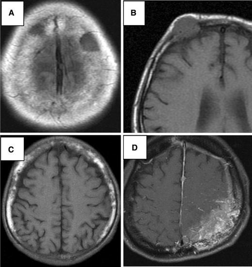
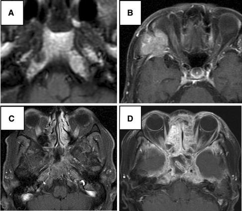
Skull metastases are malignant bone tumors which are increasing in incidence. The objectives of this study were to characterize the MR imaging features, locations, and extent of metastatic skull tumors to determine the frequency of the symptomatic disease, and to assess patient outcomes. Between September 2002 and March 2008, 175 patients undergoing routine head MR imaging were found to have metastatic skull tumors. Contrast-enhanced study with fat suppression was used in some cases when required. Classification of metastases was simplified to three yes/no questions: first, with regard to location (either in the calvarium or in the cranial base); second, with regard to distribution within the plane of the cranial bone (either "circumscribed" meaning clearly demarcated and confined to one bone, or "diffuse" and likely to spread across a suture to another bone); and third, with regard to invasion ("intraosseous" in cranial bones only, or "invasive" spreading from the skull, either out into the scalp or inward to the dura and perhaps further in). Primary sites were breast cancer (55%), lung cancer (14%), prostate cancer (6%), malignant lymphoma (5%), and others (20%). The mean time from primary diagnosis to skull metastasis diagnosis was 71 months for cases of breast cancer, 26 months for prostate cancer, 9 months for lung cancer, and 4 months for malignant lymphoma. Calvarial circumscribed intraosseous metastases were found most frequently (27%). The patients were mainly asymptomatic. However, some patients suffered from local pain or cranial nerve palsies that harmed their quality of life. Treatment, mainly for symptomatic cases, was by local or whole-skull irradiation. Metastatic skull tumors are not rare, and most are calvarial circumscribed intraosseous tumors. MR images contribute to understanding their type, location, and multiplicity, and their relationship to the brain, cranial nerves, and dural sinuses. Radiation therapy improved the QOL of patients with neurological symptoms.
Figures

References
-
- De Monte F, Hanbali F, Ballo MT. Skull base metastasis. In: Berger MS, Prados MD, editors. Text book of neuro-oncology. Philadelphia: Elsevier; 2005. pp. 466–475.
-
- Greenberg HS, Deck MD, Vikram B. Metastasis to the base of the skull: clinical findings in 43 patients. Neurology. 1981;31:530–537. - PubMed
MeSH terms
LinkOut - more resources
Full Text Sources
Medical
