. 2010 Dec;40(12):3347-57.
doi: 10.1002/eji.201041037.

IL-1β regulates a novel myeloid-derived suppressor cell subset that impairs NK cell development and function

Affiliations

IL-1β regulates a novel myeloid-derived suppressor cell subset that impairs NK cell development and function

Moshe Elkabets et al. Eur J Immunol. 2010 Dec.

Abstract

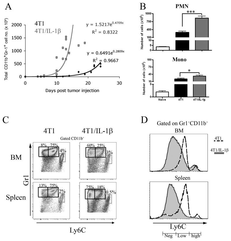
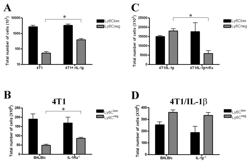
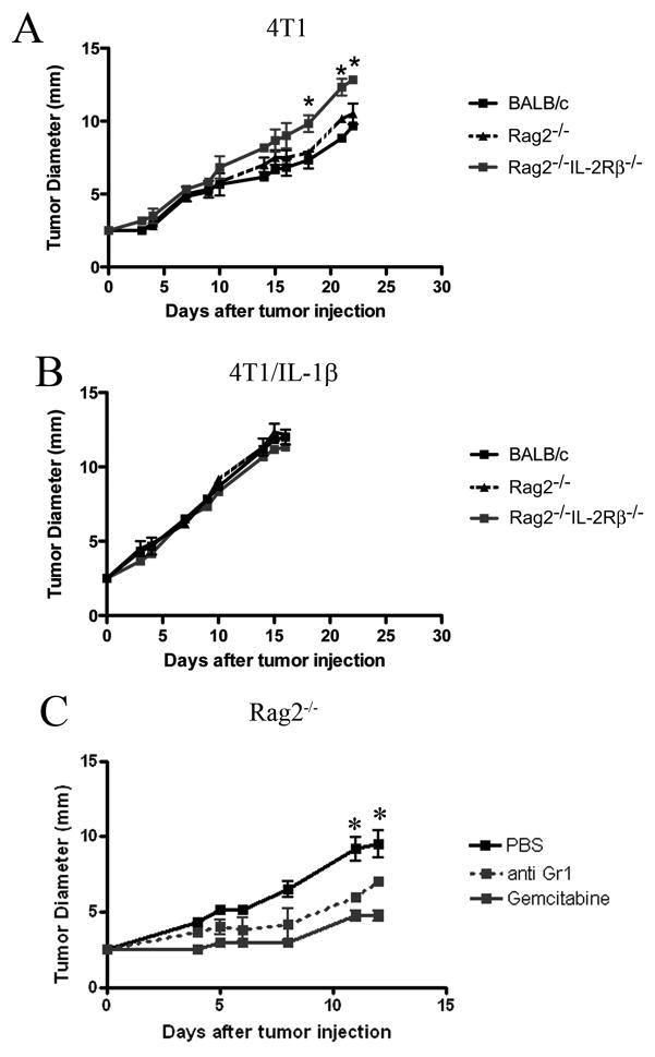
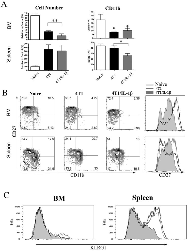
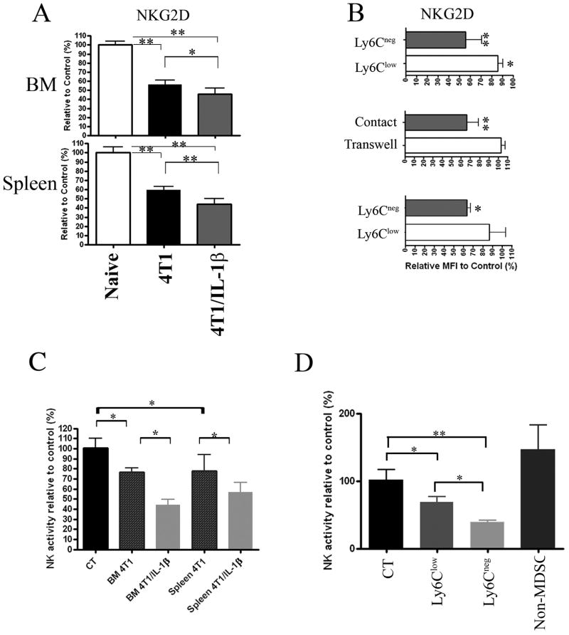
Chronic inflammation is associated with promotion of malignancy and tumor progression. Many tumors enhance the accumulation of myeloid-derived suppressor cells (MDSC), which contribute to tumor progression and growth by suppressing anti-tumor immune responses. Tumor-derived IL-1β secreted into the tumor microenvironment has been shown to induce the accumulation of MDSC possessing an enhanced capacity to suppress T cells. In this study, we found that the enhanced suppressive potential of IL-1β-induced MDSC was due to the activity of a novel subset of MDSC lacking Ly6C expression. This subset was present at low frequency in tumor-bearing mice in the absence of IL-1β-induced inflammation; however, under inflammatory conditions, Ly6C(neg) MDSC were predominant. Ly6C(neg) MDSC impaired NK cell development and functions in vitro and in vivo. These results identify a novel IL-1β-induced subset of MDSC with unique functional properties. Ly6C(neg) MDSC mediating NK cell suppression may thus represent useful targets for therapeutic interventions.


Conflict of interest statement

Conflict of interest

The authors declare no financial or commercial conflict of interest.

Figures

Figure 1
Figure 1. 4T1/IL-1β tumors enhance the accumulation of splenic Ly6Cneg MDSC
A) 2 × 10⁵ 4T1 and 4T1/IL-1β tumor cells, respectively, were injected into the right footpad of BALB/c mice. Spleen cells from tumor-bearing mice (n=12–16) were counted and single cell suspensions were stained for FACS analysis. Total Gr-1+CD11b+ (black line - 4T1, grey line -4T1/IL-1β) cell numbers were calculated at the indicated days after tumor cell injection. Regression curves were calculated using MS Excel. CD11b+Gr-1+ cells were identified as indicated in Supplemental Figure 1. (B–D) Splenocytes from tumor-bearing BALB/c mice (tumor diameter: 10–12 mm) were stained for Gr-1, CD11b, Ly6C. (B) Total numbers of PMN-MDSC and Mono-MDSC present in the different tumor-bearing mice. (C) Gr-1 versus Ly6C expression on gated CD11b+ cells. CD11b+ cells were identified as indicated in Supplemental Figure 2A. (D) Histograms of Ly6C expression by Gr-1+CD11b+ cells from the indicated tumor bearing mice. (Shaded–4T1, dotted line - 4T1/IL-1β). The results show mean values pooled from 3–5 independent experiments (n=3–5 for each experiment). Error bars show SD. **p<0.01, *p<0.05, calculated using the two-sided Student’s t-test.
Figure 2
Figure 2. Tumor and host derived IL-1β regulates size of splenic Ly6Cneg MDSC pool
Cells were identified as indicated in Figure 1B and Supplemental Figure 2A. (A) The total numbers of PMN-MDSC isolated from BALB/c mice bearing 4T1 (tumor diameter: 7 mm) treated with or without rIL-1β. (B) The total numbers of PMN-MDSC isolated from BALB/c and IL-1Ra−/− mice bearing 4T1. (C) The total numbers of PMN-MDSC isolated from BALB/c mice bearing 4T1/IL-1β tumor cells (tumor diameter: 7 mm) treated with or without rIL-1Ra. (D) The total numbers of PMN-MDSC isolated from BALB/c and IL-1β−/− mice bearing 4T1/IL-1β tumor cells. Error bars show SD. *p<0.05, calculated two-sided Student’s t-test.
Figure 3
Figure 3. Enhanced tumorigenicity of IL-1β-secreting 4T1 tumors
Tumor diameters at the indicated time points after injection of 2×10⁵ (A) 4T1 and (B) 4T1/IL-1β tumor cells into the footpad of BALB/c, Rag2−/− and Rag−/−IL-2Rβ−/− mice (n=3–4). (C) 4T1/IL-1β tumor cells were injected into the footpad of Rag2−/− mice (n=4); MDSC were eliminated by Gemcitabine treatment or depleted with antibodies against Gr-1. Tumor diameters were measured every 2–3 days. One representative experiment from 2–3 preformed is shown. Error bars show SD. *p<0.05, calculated two-sided Student’s t-test.
Figure 4
Figure 4. Impaired NK cell development in 4T1/IL-1β tumor bearing mice
Cells of BM (top) and spleen (bottom) from naïve Rag2−/− and Rag2−/− mice bearing 4T1 or 4T1/IL-1β tumors. NK cells were identified as shown in Supplemental Figure 4. (A) Left, Comparison of CD122+ NKP46+ NK cell numbers from the indicated tumor bearing mice to naïve controls (controls were set to 100%). Right, percentage of CD11b+ cells among CD122+NKp46+ cells. (B) Contour plot of CD27 versus CD11b expression and histogram of CD27 expression on NK cells. (C) Histogram of KLRG1 expression on BM ‘left) and splenic(right) CD122+NKp46+ NK cells. All data are derived from 6–8 mice bearing tumors of 9–11mm diameter in 3 independent experiments. Error bars show SD. **p<0.01, *p<0.05 using two-sided Student’s t-test.
Figure 5
Figure 5. MDSC from tumor-bearing mice affect NKG2D expression by NK cells
NK cells were identified as indicated in Supplemental Figure 4. (A) Mean fluorescence intensity of NKG2D expression by BM (top) and splenic (bottom) CD122+NKp46+ NK cells from Rag2−/−mice bearing 4T1 and 4T1/IL-1β tumors (n=6–8), respectively. Results are shown relative to NK cells from naïve Rag2−/− (controls were set to 100%). One representative result from 3 independent experiments is shown. (B) NKG2D expression by CD122+NKP46+DX5+ cells among Rag2−/− splenocytes after 24h-co-culture with enriched Ly6Clow and Ly6Cneg MDSC (top), or after 24h-co-culture of naïve Rag2−/− splenocytes with enriched Ly6Cneg MDSC in transwell plates (middle). NKG2D expression by splenic NK cells (CD122+DX5+NKP46+) 3 days after adoptive transfer of enriched Ly6Cneg and Ly6Clow MDSC, respectively, into naïve Rag2−/− mice (bottom). Results from one representative experiment of three performed. (C) Activity of host NK cells measured in vivo after i.v. injection of 2x10⁶ MACS-enriched Gr-1+CD11b+ cells from BM or spleen of mice bearing 4T1 and 4T1/IL-1β tumors as indicated. Activity is given relative to NK cells from non-injected control mice (n=4–5). Results from one representative experiment of two performed. (D) Ly6Clow MDSC, Ly6Cneg MDSC and non-MDSC were sorted from spleens of 4T1/IL-1β tumor-bearing mice and 5x10⁵ cells of each population were injected i.v. into separate naïve BALB/c mice (n=5) and NK cell activity was measured in vivo. Results from one representative experiment of two performed. Error bars show SD. **p<0.01, *p<0.05 using two-sided Student’s t-test.

Comment in

Similar articles

Cited by

References

    1. Mantovani A, Allavena P, Sica A, Balkwill F. Cancer-related inflammation. Nature. 2008;454:436–444. - PubMed
    1. Lin WW, Karin M. A cytokine-mediated link between innate immunity, inflammation, and cancer. J Clin Invest. 2007;117:1175–1183. - PMC - PubMed
    1. Apte RN, Voronov E. Is interleukin-1 a good or bad ‘guy’ in tumor immunobiology and immunotherapy? Imunol Rev. 2008;222:222–241. - PubMed
    1. Dinarello CA. Immunological and inflammatory functions of the interleukin-1 family. Annu Rev Immunol. 2009;27:519–550. - PubMed
    1. Voronov E, Shouval DS, Krelin Y, Cagnano E, Benharroch D, Iwakura Y, Dinarello CA, Apte RN. IL-1 is required for tumor invasiveness and angiogenesis. Proc Natl Acad Sci U S A. 2003;100:2645–2650. - PMC - PubMed

Publication types

MeSH terms