Review
. 2011 Jan;33(1):61-72.
doi: 10.1002/bies.201000095.

Identifying roles for neurotransmission in circuit assembly: insights gained from multiple model systems and experimental approaches

Affiliations
Review

Identifying roles for neurotransmission in circuit assembly: insights gained from multiple model systems and experimental approaches

Adam Bleckert et al. Bioessays. 2011 Jan.

Abstract

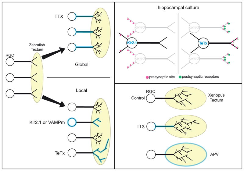
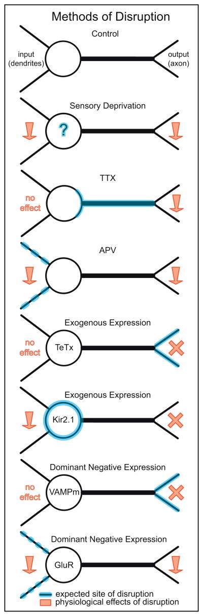
In the adult nervous system, chemical neurotransmission between neurons is essential for information processing. However, neurotransmission is also important for patterning circuits during development, but its precise roles have yet to be identified, and some remain highly debated. Here, we highlight viewpoints that have come to be widely accepted or still challenged. We discuss how distinct techniques and model systems employed to probe the developmental role of neurotransmission may reconcile disparate ideas. We underscore how the effects of perturbing neurotransmission during development vary with model systems, the stage of development when transmission is altered, the nature of the perturbation, and how connectivity is assessed. Based on findings in circuits with connectivity arranged in layers, we raise the possibility that there exist constraints in neuronal network design that limit the role of neurotransmission. We propose that activity-dependent mechanisms are effective in refining connectivity patterns only when inputs from different cells are close enough, spatially, to influence each other's outcome.


Figures

Box 1
Box 1
Figure 1
Figure 1. Stages of circuit development
Simplified view of the major developmental events underlying the assembly of neuronal circuits. I. Both axons and dendrites show marked motility during early stages of neuronal growth. Some sites of contact differentiate into synapses (e.g. red arrow) upon the recruitment of pre- (yellow) and postsynaptic (red) proteins. II. Many circuits undergo a period of refinement in their initial pattern of connectivity by the removal of erroneous contacts (compare with III). Individual presynaptic cells may contact more postsynaptic cells than at maturity (greater divergence), and single postsynaptic cells may receive inputs from inappropriate presynaptic cells (greater convergence). Note that synapse elimination (blue arrow) and synapse formation (red arrow) can take place concurrently. III. Mature patterns of circuits are established not only by synapse elimination but also by the subsequent growth and maintenance of appropriate connections.
Figure 2
Figure 2. Circuits that refine their connectivity based on neurotransmission
In many systems, postsynaptic cells receive erroneous connections that are eliminated by maturity. A: At the mammalian neuromuscular junction (NMJ), multiple motor neurons (MN) contact a muscle fiber (MF) at a single junction in early development but only one axon remains at maturity. B: In some vertebrate visual systems, connections from retinal ganglion cells (RGC) representing the left and right eyes are segregated into eye-specific layers in the dorsal lateral geniculate nucleus (dLGN) and ocular dominance columns (ODCs) in the visual cortex (VC). Eye-specific layers form prior to eye-opening and before ODCs appear. C: In the rodent cerebellum, multiple cells from the inferior olivary nucleus make climbing fiber (CF) connections onto the cell body of each Purkinje cell (PC). All but one input is subsequently removed, and the remaining climbing fiber expands its territory to innervate the proximal dendrites of the Purkinje cell.
Figure 3
Figure 3. Methodologies for disrupting neurotransmission
Summary of common methods used to disrupt neurotransmission. Some approaches suppress transmission onto a cell (input), perturb transmission from the cell (output) or affect both input and output. Methods that suppress release of transmitters include the use of Tetanus toxin, TeTx; Dominant negative expression of mutated vesicle associated membrane protein, VAMPm [91], and Tetrodotoxin (TTX). Sensory Deprivation, N-methyl-D-aspartate (NMDA) receptor blocker (APV) and over-expression of the inward rectifying potassium channel Kir2.1 or nonfunctioning forms of postsynaptic receptors (e.g. the C terminal of glutamate receptors (GluR) [48,92]) alter transmission by reducing both input and output. Downward red arrows, decrease neurotransmission; red crosses, blockade of transmission.
Figure 4
Figure 4. One type of in vivo perturbation with distinct effects across two model systems
A: In the mammalian olfactory system, subpopulations of olfactory sensory neurons (OSNs) (blue and yellow cells) express a single type of olfactory receptor. Each population projects their axons from the olfactory sensory epithelium (OSE) to the olfactory bulb (OB) where they converge at separate postsynaptic specializations or glomeruli (green and purple ovals). TeTx expression in a subpopulation (C) but not all (B) OSNs leads to mistargeting of axons [62]. D: In the vertebrate retina, ON and OFF bipolar cells (yellow) receive their inputs from photoreceptors (Ph) and contact retinal ganglion cells (RGCs). ON bipolar cells stratify their axons in the inner half of the inner plexiform layer (IPL). RGC dendrites stratify in either the ON or OFF or both sublayers (ON/OFF) of the IPL. Expression of TeTX in all ON bipolar cells does not disrupt their axonal morphology and lamination nor alter the dendritic structure of RGCs [64]. However, compared to wildtype animals (E) ON RGCs make fewer connections (cyan dots) in the TeNT retina (F).
Figure 5
Figure 5. Is neurotransmission-mediated circuit refinement dependent on input proximity?
A: Neurotransmission dependent elimination of motor neuron (MN) axonal inputs that initially overlap at the neuromuscular junction is thought to employ local ‘punishment’ signals (red arrows, a) [73]. MF, muscle fiber. B: Parallel fibers (PFs) make connections primarily onto the distal dendrites and a single climbing fiber (CF) innervates the proximal dendrites of cerebellar Purkinje cells. While climbing fibers are the first to innervate Purkinje cells, their territory becomes intermingled with the inputs of parallel fibers later in development. Disruptions to neurotransmission from either PFs or CFs result in a local take over of territory by the more active input (red arrows, b; [100]). C & D: Not all circuits with converging afferents display neurotransmission-mediated competition. C: Inputs representing the ipsilateral (ipsi) and contralateral (contra) ears contact separate dendritic arbors of Nucleus Laminaris (NL) neurons of the auditory brainstem. These inputs do not intermingle even during development. If transmission is disrupted in one set of inputs, synaptic takeover does not occur [101]. D: Similarly, in the mammalian retina, the axons of OFF bipolar cells (BCs) do not innervate the retinal ganglion cell (RGC) dendrites contacted by ON BC axons with suppressed transmitter release [64]. The apparent absence of activity-dependent mechanisms (crossed arrows in c,d) in shaping the selectivity of inputs in the NL and retina may be because distinct types of afferents in their circuits show little to no spatial overlap. The axonal arbors of neighboring BCs of the same subtype tile at maturity but whether or not these converging inputs intermingle during development, and utilize neurotransmission to determine their relative territories has yet to be determined (e, en face view of the retina).

Similar articles

Cited by

References

    1. Hubel DH, Wiesel TN, LeVay S. Plasticity of ocular dominance columns in monkey striate cortex. Philos Trans R Soc Lond B Biol Sci. 1977;278:377–409. - PubMed
    1. LeVay S, Wiesel TN, Hubel DH. The development of ocular dominance columns in normal and visually deprived monkeys. J Comp Neurol. 1980;191:1–51. - PubMed
    1. Sanes JR, Lichtman JW. Development of the vertebrate neuromuscular junction. Annu Rev Neurosci. 1999;22:389–442. - PubMed
    1. Hume RI, Role LW, Fischbach GD. Acetylcholine release from growth cones detected with patches of acetylcholine receptor-rich membranes. Nature. 1983;305:632–4. - PubMed
    1. Taylor J, Docherty M, Gordon-Weeks PR. GABAergic growth cones: release of endogenous gamma-aminobutyric acid precedes the expression of synaptic vesicle antigens. J Neurochem. 1990;54:1689–99. - PubMed

Publication types