. 2010 Nov 26:5:31.
doi: 10.1186/1750-1172-5-31.

In vivo effects of antibodies from patients with anti-NMDA receptor encephalitis: further evidence of synaptic glutamatergic dysfunction

Affiliations

In vivo effects of antibodies from patients with anti-NMDA receptor encephalitis: further evidence of synaptic glutamatergic dysfunction

Mario Manto et al. Orphanet J Rare Dis. .

Abstract

Background: A severe encephalitis that associates with auto-antibodies to the NR1 subunit of the NMDA receptor (NMDA-R) was recently reported. Patients' antibodies cause a decrease of the density of NMDA-R and synaptic mediated currents, but the in vivo effects on the extracellular glutamate and glutamatergic transmission are unknown.

Methods: We investigated the acute metabolic effects of patients' CSF and purified IgG injected in vivo. Injections were performed in CA1 area of Ammon's horn and in premotor cortex in rats.

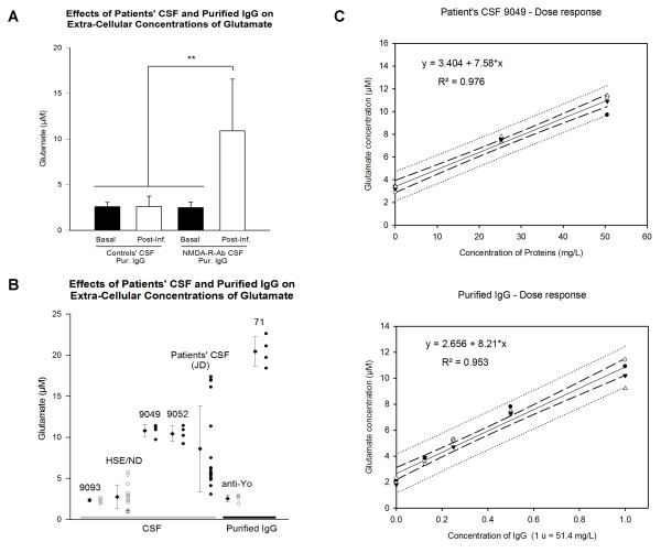
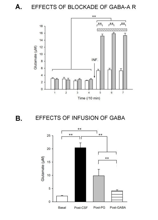
Results: Patient's CSF increased the concentrations of glutamate in the extracellular space. The increase was dose-dependent and was dramatic with purified IgG. Patients' CSF impaired both the NMDA- and the AMPA-mediated synaptic regulation of glutamate, and did not affect the glial transport of glutamate. Blockade of GABA-A receptors was associated with a marked elevation of extra-cellular levels of glutamate following a pretreatment with patients' CSF.

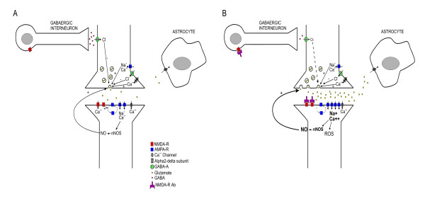
Conclusion: These results support a direct role of NMDA-R antibodies upon altering glutamatergic transmission. Furthermore, we provide additional evidence in vivo that NMDA-R antibodies deregulate the glutamatergic pathways and that the encephalitis associated with these antibodies is an auto-immune synaptic disorder.


Figures

Figure 1
Figure 1
Example of histological section at the level of CA1 area after experiment. Location of the guide (G) and tip (T) of the microdialysis probe in right CA1 of the rat after experiment. Ent: entorhinal cortex; MG: medial geniculate nuclei; MP: medial mammillary nucleus; fmj: forceps major corpus callosum.
Figure 2
Figure 2
Effects on extra-cellular concentrations of glutamate. A: comparison of the glutamate concentrations measured before (basal) and after infusion of CSF or purified IgGs of patients (Post-Inf.). Each sample (5 control CSF, 6 patients' CSF with NMDA-R-Ab, one purified IgGs positive for NMDA-R-Ab and one purified IgGs positive for anti-Yo antibodies) was infused in 4 different rats. Means and SDs are represented. Measurements were made at baseline (before infusion), and between 30 and 60 minutes after CSF or purified IgGs infusion. B: point plot (one point corresponds to one rat injected) of the extra-cellular concentrations of glutamate following infusion of CSF (open circles: controls' CSF; filled circles: patients' CSF), and purified IgGs (open circles: control IgGs from a patient with Yo-Ab; filled circles: purified IgGs from a patient positive for NMDA-R-Ab) measured in CA1 zone between 30 and 60 minutes after CSF infusion. C: Top panel: concentrations of extra-cellular glutamate according to the dilution of patients' CSF infused in the CA1 area. Four rats were infused with 3 successive doses from one patient's CSF positive for NMDA-R-Ab (CSF 9049). Linear regression, 95% confidence intervals (dashed lines) and 95% prediction intervals (dotted lines) are shown. Bottom panel: Similar results were obtained with purified IgG of CSF 9052. Four rats were individually infused with 4 successive doses. **: p < 0.01.
Figure 3
Figure 3
Effects of patients' CSF on glutamate receptor functions. A: Effects of infusion of patients' CSF on the NMDA-mediated regulation of glutamate. Groups 1 and 2: n = 8 rats injected with 5 control CSF. Groups 3 and 4: n = 8 rats injected with CSF from 5 patients positive for NMDA-R-Ab. Groups 5 and 6: 4 rats injected with a control CSF and 4 rats injected with one CSF positive for NMDA-R-Ab, respectively. Values are mean ± SEM. B: Effects of AMPA, AMPA blockade (DNQX), combination of NMDA + DNQX and combination of NMDA + AMPA following administration of NMDA-R-Ab. Groups 1, 3 and 5: 4 rats injected with 4 control CSF. Groups 2, 4 and 6: 4 rats injected with CSF from 4 patients with NMDA-R-Ab. Group 7: 4 rats injected with 1 control CSF. Group 8: 4 rats injected with 1 CSF with NMDA-R-Ab. Values are mean ± SEM. *: p < 0.05; **: p < 0.01
Figure 4
Figure 4
Interaction with GABA-A receptors and infusion of GABA. A: Effects of blockade of GABA-A receptors on glutamate levels following infusion of patients' CSF. In rats infused (INF) with CSF with NMDA-R-Ab (9049; n = 3 rats; grey bars), administration of bicuculline (represented by a hatched rectangle) increased markedly the concentrations of glutamate. The bicuculline-induced increase of glutamate was lower in rats injected with the control solution 9093 (n = 3 rats; white bars). B: Effects of successive administration of pregabalin (PG) and GABA after infusion of purified IgGs of a patient with NMDA-R-Ab (n = 4 rats). Concentrations of glutamate raised markedly with purified IgGs. Basal: T0 min; Post-NMDA-R-Ab (Post-CSF): post-injection at T30 min; Post-PGB: T90 min post-infusion; Post-Gaba: Post-infusion of Gaba 50 μM at T120 min. Values are mean ± SD. **: p < 0.01
Figure 5
Figure 5
Effects of patient's CSF on concentrations of nitric oxide (NO). Inhibition of nNOS with 7-nitroindazole (7-NI) counteracts the effects of NMDA-R Ab. N = 4 rats infused with control CSF 9093 and n = 4 rats infused with a CSF with NMDA-R-Ab (9049). Values are mean ± SD and are expressed as percentages of baseline measurements. **: p < 0.01
Figure 6
Figure 6
Proposed scheme of the synaptic consequences of NMDA-R-Ab. The antibodies block the NR1/NR2 heteromers of the NMDA receptor, causing a state of imbalance between NMDA and AMPA receptors. Blockade of NMDA-R impairs AMPA-R endocytosis. NMDA-R-Ab cause an increased release of glutamate which is dependent on the alpha2-delta subunit of VGCC and which is also related to a deregulation of gabaergic interneurons. NO acts as a retrograde messenger and amplifies glutamate release.

References

    1. Dalmau J, Gleichman AJ, Hughes EG, Rossi JE, Peng X, Lai M, Dessain SK, Rosenfeld MR, Balice-Gordon R, Lynch DR. Anti-NMDA-receptor encephalitis: case series and analysis of the effects of antibodies. Lancet Neurol. 2008;7:1091–8. doi: 10.1016/S1474-4422(08)70224-2. - DOI - PMC - PubMed
    1. Dalmau J, Tuzun E, Wu HY, Masjuan J, Rossi JE, Voloschin A, Baehring JM, Shimazaki H, Koide R, King D, Mason W, Sansing LH, Dichter MA, Rosenfeld MR, Lynch DR. Paraneoplastic anti-N-methyl-D-aspartate receptor encephalitis associated with ovarian teratoma. Ann Neurol. 2007;61:25–36. doi: 10.1002/ana.21050. - DOI - PMC - PubMed
    1. Seki M, Suzuki S, Iizuka T, Shimizu T, Nihei Y, Suzuki N, Dalmau J. Neurological response to early removal of ovarian teratoma in anti-NMDA-R encephalitis. J Neurol Neurosurg Psychiatry. 2008;79:324–326. doi: 10.1136/jnnp.2007.136473. - DOI - PMC - PubMed
    1. Hughes EG, Peng X, Gleichman AJ, Lai M, Zhou L, Tsou R, Parsons TD, Lynch DR, Dalmau J, Balice-Gordon RJ. Cellular and synaptic mechanisms of anti-NMDA receptor encephalitis. J Neurosci. 2010;30:5866–75. doi: 10.1523/JNEUROSCI.0167-10.2010. - DOI - PMC - PubMed
    1. Manto M, Laute MA, Aguera M, Rogemond V, Pandolfo M, Honnorat J. Effects of anti-glutamic acid decarboxylase antibodies associated with neurological diseases. Ann Neurol. 2007;61:544–551. doi: 10.1002/ana.21123. - DOI - PubMed

Publication types

MeSH terms