. 2011 Jan 7;404(1):195-200.
doi: 10.1016/j.bbrc.2010.11.092. Epub 2010 Nov 24.

THAP5 is a DNA-binding transcriptional repressor that is regulated in melanoma cells during DNA damage-induced cell death

Affiliations

THAP5 is a DNA-binding transcriptional repressor that is regulated in melanoma cells during DNA damage-induced cell death

Meenakshi P Balakrishnan et al. Biochem Biophys Res Commun. .

Abstract

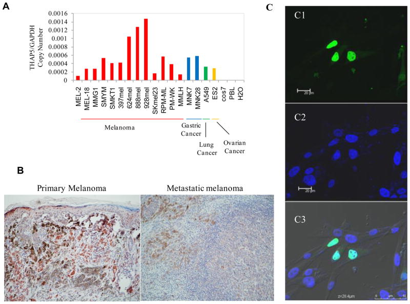
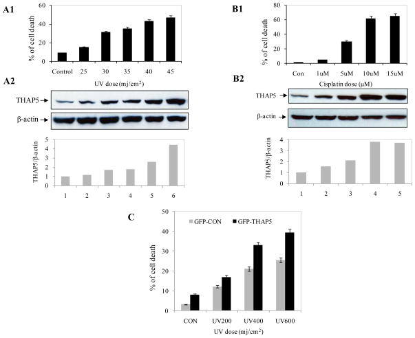
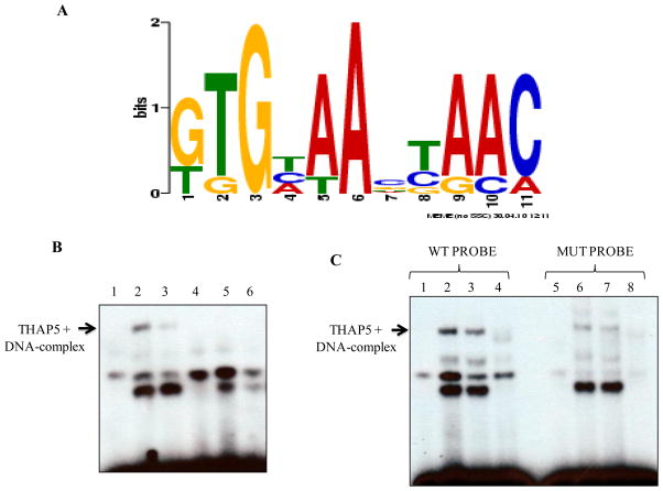
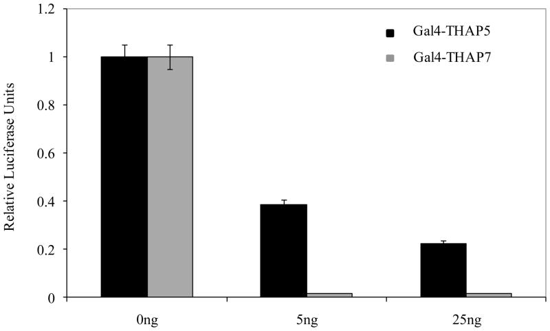
THAP5 was originally isolated as a specific interactor and substrate of the mitochondrial pro-apoptotic Omi/HtrA2 protease. It is a human zinc finger protein characterized by a restricted pattern of expression and the lack of orthologs in mouse and rat. The biological function of THAP5 is unknown but our previous studies suggest it could regulate G2/M transition in kidney cells and could be involved in human cardiomyocyte cell death associated with coronary artery disease (CAD). In this report, we expanded our studies on the properties and function of THAP5 in human melanoma cells. THAP5 was expressed in primary human melanocytes as well as in all melanoma cell lines that were tested. THAP5 protein level was significantly induced by UV irradiation or cisplatin treatment, conditions known to cause DNA damage. The induction of THAP5 correlated with a significant increase in apoptotic cell death. In addition, we show that THAP5 is a nuclear protein that could recognize and bind a specific DNA motif. THAP5 could also repress the transcription of a reporter gene in a heterologous system. Our work suggests that THAP5 is a DNA-binding protein and a transcriptional repressor. Furthermore, THAP5 has a pro-apoptotic function and it was induced in melanoma cells under conditions that promoted cell death.


Figures

Fig. 1
Fig. 1
Expression and localization of THAP5 in melanoma cells. A, THAP5 mRNA expression in various cell lines. THAP5 is expressed at various levels in all human melanoma cell lines tested. THAP5 expression was also detected in some human gastric, lung and ovarian cell lines but not in Cos7 or PBL cells. B, Imunohistochemistry of primary and metastatic melanoma tissues showing distinct THAP5 staining of melanocytes. C, Subcellular localization of the GFP-THAP5 in MeWo cells. Confocal images of MeWo cells transfected with GFP-THAP51-395 shows nuclear localization (green in C1). Panel C2 shows cells with DAPI staining for the nucleus and C3 shows merged image of Panels C1, C2 and C3.
Fig. 2
Fig. 2
THAP5 is induced following UV or cisplatin treatment. MeWo cells were treated with increasing doses of UV and cisplatin and apoptosis monitored by flow cytometry as described in the Methods (A1, B1). Extracts were prepared from the same cell populations and subjected to SDS-PAGE and Western blot analysis using THAP5 antibody. THAP5 was significantly induced with increasing doses of UV and cisplatin and this corresponds to an increased apoptosis in the treated cells (A2, B2). β-actin antibody was used to verify that equal amount of protein was present in each lane. Bottom panels, A2 and B2 show densitometry analysis. C. MeWo cells were transfected with GFPC vector and GFPC-THAP5 plasmids. Transfected cells were exposed to UV and cell death was monitored. Data are mean ± SD of 3 different experiments.
Fig. 3
Fig. 3
THAP5 recognized a specific DNA sequence. A, Identification of a consensus DNA-binding site for THAP5. This consensus sequence was created from a pool of 25 independent THAP5 bound oligonucleotides recovered at the end of 7 rounds of selection. The DNA sequences were analyzed by the motif-discovery program MEME. B, Electrophoretic Mobility Shift Assay showing THAP5 binding to the consensus DNA sequence. Lane 1, control (no extract); lane 2, MeWo nuclear extract; lane 3, MeWo extract with anti-THAP5 antibody; lane 4, NIH 3T3 extract; lane 5, MeWo extract plus 100 molar excess of cold THAP5 specific probe (WT) and lane 6, MeWo extract plus 100 molar excess of mutant probe incubated with 32P labeled wild type probe. C, Specificity of THAP5 Binding. First four lanes show THAP5 binding to the labeled WT probe. Lane 1, control (no extract); lane 2, MeWo nuclear extract; lane 3, MeWo extract with anti-THAP5 antibody; lane 4, NIH 3T3 extract. Lanes 5–6 show reduced binding of THAP5 to the labeled Mutant probe. Lane 5, control (no extract); lane 6, MeWo nuclear extract; lane 7, MeWo extract with anti-THAP5 antibody; lane 8, NIH 3T3 extract.
Fig. 4
Fig. 4
THAP5 is a transcriptional repressor. Different amounts of Gal4-THAP5 or Gal4-THAP7 vectors were transfected into HEK293T cells along with a Gal4-luciferase reporter. Luciferase assays were performed 48 hrs post transfection. Gal4-THAP5 was able to repress the transcription of the reporter gene but to a lesser extent than the Gal4-THAP7. Data are means ± SD of four independent experiments.

References

    1. Roussigne M, Kossida S, Lavigne AC, Clouaire T, Ecochard V, Glories A, Amalric F, Girard JP. The THAP domain: a novel protein motif with similarity to the DNA-binding domain of P element transposase. Trends Biochem Sci. 2003;28:66–69. - PubMed
    1. Cayrol C, Lacroix C, Mathe C, Ecochard V, Ceribelli M, Loreau E, Lazar V, Dessen P, Mantovani R, Aguilar L, Girard JP. The THAP-zinc finger protein THAP1 regulates endothelial cell proliferation through modulation of pRB/E2F cell-cycle target genes. Blood. 2007;109:584–594. - PubMed
    1. Clouaire T, Roussigne M, Ecochard V, Mathe C, Amalric F, Girard JP. The THAP domain of THAP1 is a large C2CH module with zinc-dependent sequence-specific DNA-binding activity. Proc Natl Acad Sci U S A. 2005;102:6907–6912. - PMC - PubMed
    1. Deiss LP, Feinstein E, Berissi H, Cohen O, Kimchi A. Identification of a novel serine/threonine kinase and a novel 15-kD protein as potential mediators of the gamma interferon-induced cell death. Genes Dev. 1995;9:15–30. - PubMed
    1. Gale M, Jr, Blakely CM, Hopkins DA, Melville MW, Wambach M, Romano PR, Katze MG. Regulation of interferon-induced protein kinase PKR: modulation of P58IPK inhibitory function by a novel protein, P52rIPK. Mol Cell Biol. 1998;18:859–871. - PMC - PubMed

Publication types