Review
. 2011 Apr;274(1-2):129-41.
doi: 10.1016/j.heares.2010.11.001. Epub 2010 Nov 24.

VGLUT1 and VGLUT2 mRNA expression in the primate auditory pathway

Affiliations
Review

VGLUT1 and VGLUT2 mRNA expression in the primate auditory pathway

Troy A Hackett et al. Hear Res. 2011 Apr.

Abstract

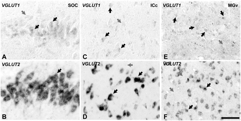
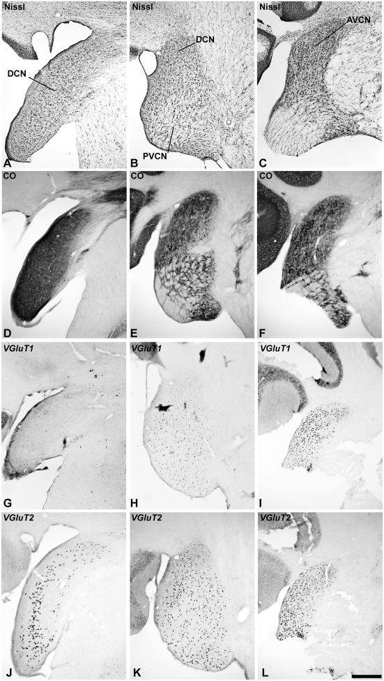
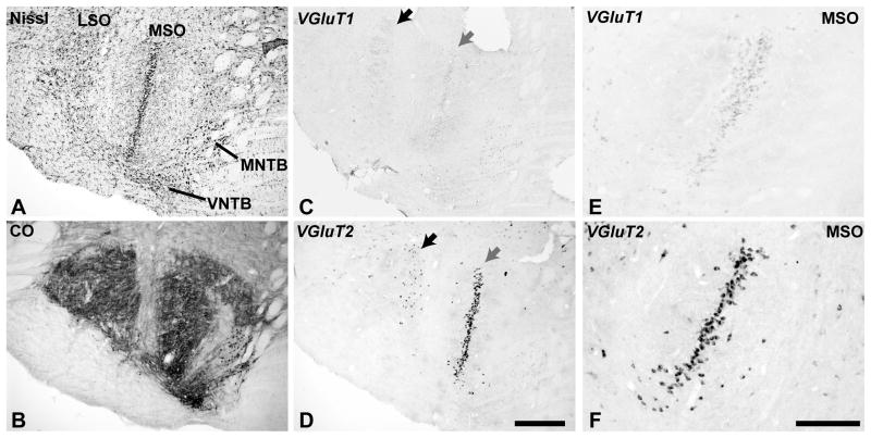
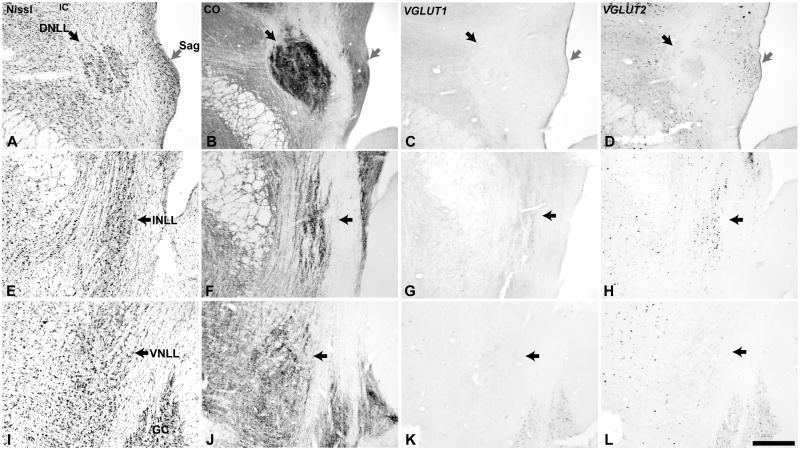
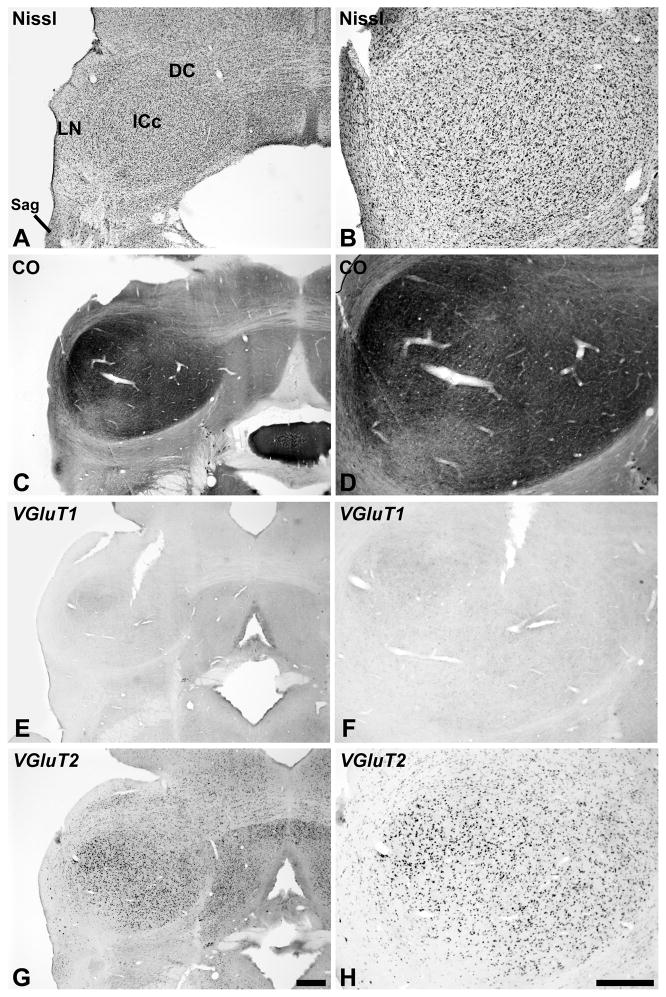
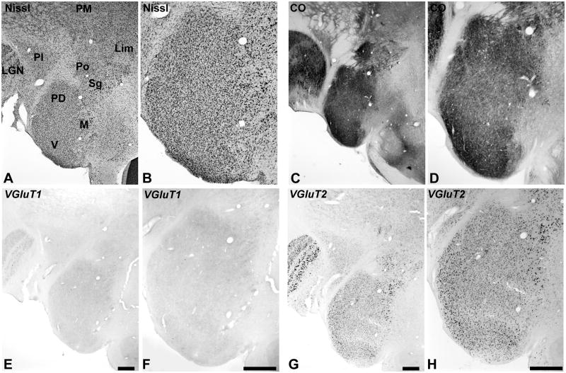
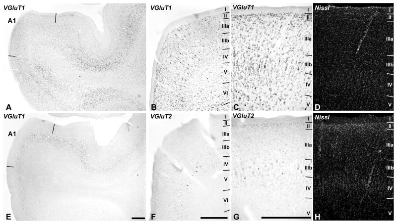
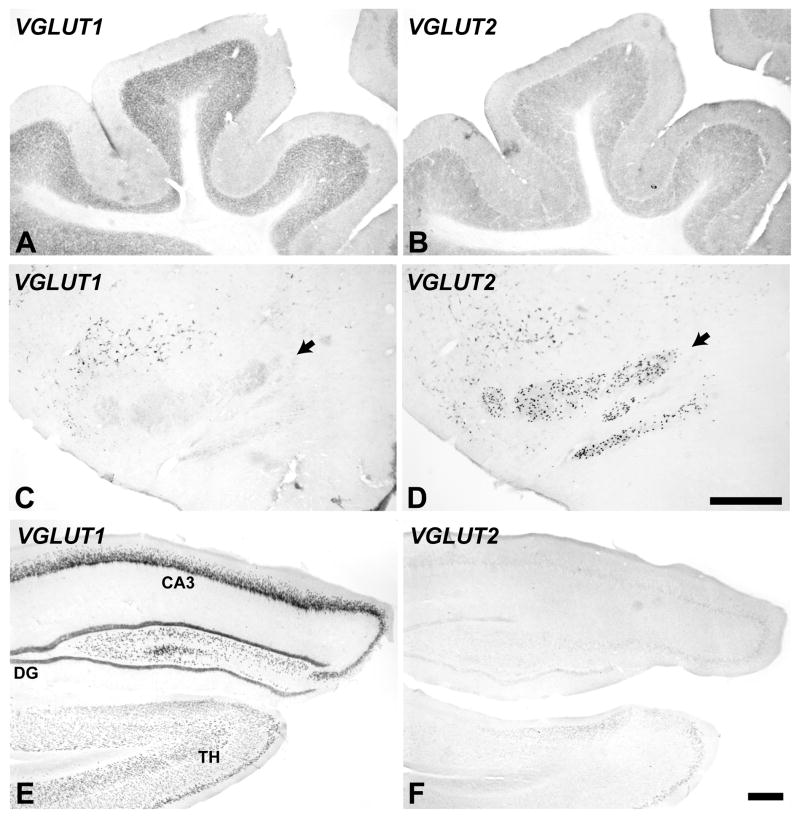
The vesicular glutamate transporters (VGLUTs) regulate the storage and release of glutamate in the brain. In adult animals, the VGLUT1 and VGLUT2 isoforms are widely expressed and differentially distributed, suggesting that neural circuits exhibit distinct modes of glutamate regulation. Studies in rodents suggest that VGLUT1 and VGLUT2 mRNA expression patterns are partly complementary, with VGLUT1 expressed at higher levels in the cortex and VGLUT2 prominent subcortically, but with overlapping distributions in some nuclei. In primates, VGLUT gene expression has not been previously studied in any part of the brain. The purposes of the present study were to document the regional expression of VGLUT1 and VGLUT2 mRNA in the auditory pathway through A1 in the cortex, and to determine whether their distributions are comparable to rodents. In situ hybridization with antisense riboprobes revealed that VGLUT2 was strongly expressed by neurons in the cerebellum and most major auditory nuclei, including the dorsal and ventral cochlear nuclei, medial and lateral superior olivary nuclei, central nucleus of the inferior colliculus, sagulum, and all divisions of the medial geniculate. VGLUT1 was densely expressed in the hippocampus and ventral cochlear nuclei, and at reduced levels in other auditory nuclei. In the auditory cortex, neurons expressing VGLUT1 were widely distributed in layers II-VI of the core, belt and parabelt regions. VGLUT2 was expressed most strongly by neurons in layers IIIb and IV, weakly by neurons in layers II-IIIa, and at very low levels in layers V-VI. The findings indicate that VGLUT2 is strongly expressed by neurons at all levels of the subcortical auditory pathway, and by neurons in the middle layers of the cortex, whereas VGLUT1 is strongly expressed by most if not all glutamatergic neurons in the auditory cortex and at variable levels among auditory subcortical nuclei. These patterns imply that VGLUT2 is the main vesicular glutamate transporter in subcortical and thalamocortical (TC) circuits, whereas VGLUT1 is dominant in corticocortical (CC) and corticothalamic (CT) systems of projections. The results also suggest that VGLUT mRNA expression patterns in primates are similar to rodents, and establish a baseline for detailed studies of these transporters in selected circuits of the auditory system.


Figures

Fig. 1
Fig. 1
Low magnification images of the owl monkey brainstem and thalamus at the level of the cochlear nucleus (left), inferior colliculus and superior olivary complex (middle), and medial geniculate (right). At each level, VGLUT1 and VGLUT2 mRNA expression are illustrated in adjacent sections to show their respective distribution patterns. Scale bar = 1mm.
Fig. 2
Fig. 2
High magnification images of cells containing VGLUT1 and VGLUT2 mRNA. VGLUT1 signals (A, C, E) were found at reduced levels in these subcortical auditory structures, and its subcellular distribution was partial compared to VGLUT2 (B, D, F). Medial superior olivary nucleus (A, B); central nucleus of the inferior colliculus (C, D); ventral division of the medial geniculate (E, F). Scale bar = 50 um.
Fig. 3
Fig. 3
VGLUT mRNA expression in the cochlear nucleus (CN). Adjacent series of sections of the cochlear nucleus at the level of the DCN (left columns); PVCN (middle columns), and AVCN (right columns). Sections illustrated at each level were processed for Nissl (A – C), cytochrome oxidase (CO) (D – F), VGLUT1 mRNA (G – I), and VGLUT2 mRNA (J – L) by in situ hybridization. Scale bars = 500 um.
Fig. 4
Fig. 4
VGLUT mRNA expression in the superior olivary complex (SOC). Adjacent sections processed for Nissl (A), CO (B), VGLUT1 mRNA (C, D) and VGLUT2 mRNA (E, F). MSO, medial superior olive; LSO, lateral superior olive; MNTB and LNTB, medial and lateral nuclei of the trapezoid body. Scale bars = 500 um.
Fig. 5
Fig. 5
VGLUT mRNA expression in the lateral lemniscus (LL) and sagulum (Sag). Adjacent sections through the dorsal nucleus (DNLL, black arrow) and sagulum (gray arrow)(A – D), intermediate nucleus (INLL, black arrow)(E – H), and ventral nucleus (VNLL, black arrow)(I – L). Sections illustrated at each level were processed for Nissl (A, E, I), cytochrome oxidase (CO) (B, F, J), VGLUT1 mRNA (C, G, K), and VGLUT2 mRNA (D, H, L) by in situ hybridization. GC, substantia griseum centralis. Scale bars = 500 um.
Fig. 6
Fig. 6
VGLUT mRNA expression in the inferior colliculus (IC). The general locations of the central (ICc), lateral (LN), dorsal cortex (DC) divisions are indicted in panel A. Sections processed for Nissl (A, B), CO (C, D), VGLUT1 mRNA (E, F) and VGLUT2 mRNA (G, H) are illustrated at two different magnifications to show detail. Scale bars = 500 um.
Fig. 7
Fig. 7
VGLUT mRNA expression in the medial geniculate complex (MGC) and adjoining nuclei of the posterior thalamus. The posterodorsal (PD), medial or magnocellular (M), and ventral (V) divisions of the MGC are indicated in panel A, stained for Nissl. Also indicated are the locations of the lateral geniculate (LGN), inferior pulvinar (PI), medial pulvinar (PM), posterior (Po), suprageniculate (Sg), and limitans (Lim) nuclei. CO expression illustrated in panels C and D. VGLUT1 mRNA (E, F). VGLUT2 mRNA (G, H), shown at different magnifications. Scale bars = 500 um.
Fig. 8
Fig. 8
VGLUT1 and VGLUT2 mRNA expression in auditory cortex. VGLUT1 mRNA (A – C) is expressed in neurons spanning layers II – VI. VGLUT2 mRNA (E – G) expression is strongest in layer IIIb, weaker in layers II, IIIa, and IV, and very low or absent in layers V and VI. Paired panels (C – D) and (G – H) are images of the same sections through A1, dual-labeled for mRNA by in situ hybridization (C, G) and fluorescent Nissl stain (D, H) to show laminar details. Scale bars = 500 um.
Fig. 9
Fig. 9
VGLUT mRNA expression in the cerebellum (A, B), inferior olivary complex (C, D black arrows), and hippocampus (E, F). VGLUT1 mRNA (left panels). VGLUT2 mRNA (right panels). Scale bars = 500 um.

References

    1. Adams JC, Mugnaini E. Dorsal nucleus of the lateral lemniscus: a nucleus of GABAergic projection neurons. Brain Res Bull. 1984;13:585–90. - PubMed
    1. Altschuler RA, Tong L, Holt AG, Oliver DL. Immunolocalization of vesicular glutamate transporters 1 and 2 in the rat inferior colliculus. Neuroscience. 2008;154:226–32. - PMC - PubMed
    1. Aoki E, Semba R, Keino H, Kato K, Kashiwamata S. Glycine-like immunoreactivity in the rat auditory pathway. Brain Res. 1988;442:63–71. - PubMed
    1. Barroso-Chinea P, Castle M, Aymerich MS, Lanciego JL. Expression of vesicular glutamate transporters 1 and 2 in the cells of origin of the rat thalamostriatal pathway. J Chem Neuroanat. 2008;35:101–7. - PubMed
    1. Barroso-Chinea P, Castle M, Aymerich MS, Perez-Manso M, Erro E, Tunon T, Lanciego JL. Expression of the mRNAs encoding for the vesicular glutamate transporters 1 and 2 in the rat thalamus. J Comp Neurol. 2007;501:703–15. - PubMed

Publication types

MeSH terms

Substances