. 2010 Nov 24;143(5):789-801.
doi: 10.1016/j.cell.2010.11.001.

Abortive HIV infection mediates CD4 T cell depletion and inflammation in human lymphoid tissue

Affiliations

Abortive HIV infection mediates CD4 T cell depletion and inflammation in human lymphoid tissue

Gilad Doitsh et al. Cell. .

Erratum in

  • Cell. 2014 Feb 27;156(5):1112-3

Abstract

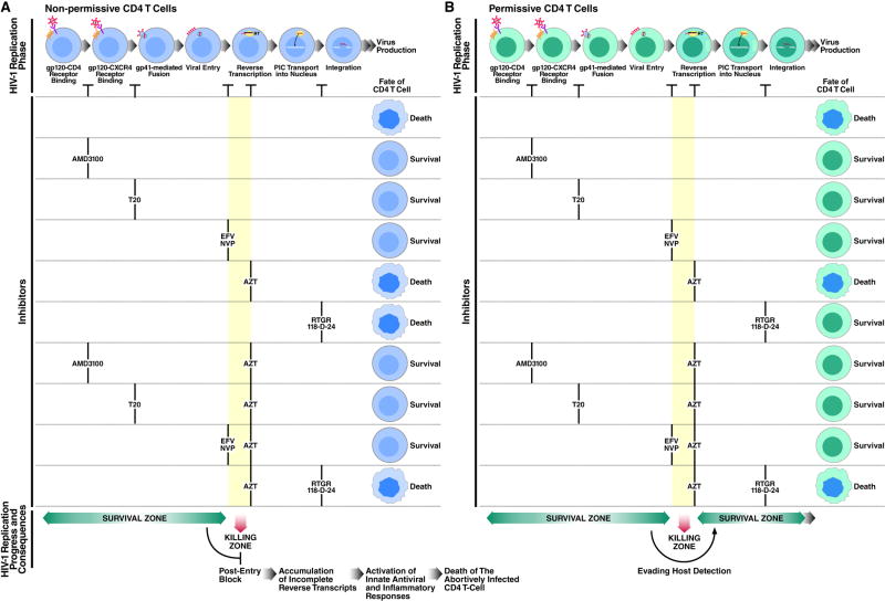
The mechanism by which CD4 T cells are depleted in HIV-infected hosts remains poorly understood. In ex vivo cultures of human tonsil tissue, CD4 T cells undergo a pronounced cytopathic response following HIV infection. Strikingly, >95% of these dying cells are not productively infected but instead correspond to bystander cells. We now show that the death of these "bystander" cells involves abortive HIV infection. Inhibitors blocking HIV entry or early steps of reverse transcription prevent CD4 T cell death while inhibition of later events in the viral life cycle does not. We demonstrate that the nonpermissive state exhibited by the majority of resting CD4 tonsil T cells leads to accumulation of incomplete reverse transcripts. These cytoplasmic nucleic acids activate a host defense program that elicits a coordinated proapoptotic and proinflammatory response involving caspase-3 and caspase-1 activation. While this response likely evolved to protect the host, it centrally contributes to the immunopathogenic effects of HIV.


Figures

Figure 1
Figure 1. Massive Depletion of CD4 T Cells in HLACs Containing Small Number of Productively Infected Cells
(A) Kinetics of spreading viral infection versus depletion of CD4 T cells after infection of HLACs with a replication-competent HIV reporter virus encoding GFP. CD4 downregulation in GFP-positive cells likely represents the combined action of the HIV Nef, Vpu, and Env proteins expressed by this virus. Ratios of viable CD4 versus CD8 T cells in HIV-infected and uninfected cultures are also shown. Flow cytometry plots represent live-gated cells, based on the forward-scatter versus side-scatter profile of the complete culture. These data are the representative results of six independent experiments utilizing tonsil cells from six different donors.
Figure 2
Figure 2. CD4 T-cell Depletion in HIV-1-Infected HLACs Predominantly Involves Non-productively Infected Cells
(A) Experimental strategy to assess indirect cell killing in HIV-1-infected human lymphoid cultures. Fresh human tonsil tissue from a single donor is processed into HLAC, and then separated into two fractions. One fraction is challenged with HIV-1 and cultured for 6 days, allowing viral spread. On day 5, the uninfected fraction is treated with AZT (5 μM) and labeled with CFSE (1 μM). On day 6, the infected and CFSE-labeled cultures are mixed and co-cultured in the presence of AZT. Because of its site of action, AZT does not block viral output from the HIV-infected cells but prevents productive infection of CFSE-labeled cells. After 6 days of co-culturing, the number of viable CSFE-positive cells is determined by flow cytometry. (B) Flow cytometry analysis of the mixed HLACs. Indirect killing is determined by gating on live CFSE-positive cells in the mixed cultures. Effector cells are either infected or uninfected cells. (C) Extensive depletion of non-productively infected CD4 T cells by HIV-1. CFSE-labeled cells mixed with uninfected or infected cells were cultured in the presence of 5 μM AZT alone or together with 250 nM AMD3100. Data represent live CFSE-positive cells 6 days after co-culture with infected or uninfected effector cells. The absence of productive infection in the CFSE-positive cells was confirmed by internal p24 staining and monitoring GFP expression following infection with the NLENG1 HIV-1 reporter virus (not shown). (D) Preferential depletion of non-productively infected CD4 T cells by HIV-1. The absolute numbers of viable CFSE-positive CD4 and CD8 T cells and B cells were determined. Percentages are normalized to the number of viable CFSE-positive cells co-cultured with uninfected cells in the presence of AZT, as depicted by (*). Error bars represent standard deviations of three samples from the same donor. This experiment is the representative of more than 10 independent experiments with more than 10 donors of tonsillar tissues. See also Figure S1.
Figure 3
Figure 3. HIV-1 Fusion Is Necessary to Induce Killing of Non-Productively Infected Cells
(A and C) Concentrations of T20 that block viral infection. HLACs were infected with the indicated clones of HIV-1 in the presence of the indicated concentrations of T20 or no drugs. One hour before incubation with the virus, cells were pretreated with T20 or left untreated. At 12 hours, cells were washed extensively and cultured under the same conditions. On day 9, the viral concentration was determined using a p24gag FLAQ assay. The amount of p24gag accumulated in the absence of drugs by each viral clone (A) or by SKY (C) was defined as 100%. (B and D) Effect of T20 on indirect killing. CFSE-labeled cells were co-cultured with cells infected with the indicated viral clones in the presence of 5 μM AZT and the indicated concentrations of T20. After 6 days, indirect killing in the mixed cultures was assessed. The number of viable CFSE-positive CD4 T cells co-cultured with uninfected cells in the presence of AZT was defined as 100% (not shown). To allow successful initial infection we pseudotyped the GIA-SKY mutants with the VSV-G envelope. NL4-3, WT lab-adapted virus; WEAU 16-8, primary virus; SIM, T20-resistant virus; GIA-SKY, T20-dependent virus; GIA and SKY, single-domain mutant viruses. Representative data from three independent experiments with different donors are shown. See also Figure S2.
Figure 4
Figure 4. Killing of Non-Productively Infected CD4 T Cells Requires Fusion of Virions from Nearby HIV-1-Producing Cells
(A) Supernatants from HIV-infected HLACs are less efficient at inducing indirect killing than mixing of HIV-infected and uninfected HLACs. (B) HIV-1 virions released into the medium do not participate in indirect killing. Replacing the mixed culture with fresh RPMI every 24 hours did not impair indirect killing. Challenging HLACs with supernatants containing 20-fold more histoculture-derived virions (1 μg p24/ml) than normally accumulated in mixed cultures containing infected cells (50 ng p24/ml) did not induce indirect killing. Percentages are normalized to the number of viable CFSE-positive cells depicted by (*). (C) CFSE-labeled cells are not killed when HIV-infected HLAC is physically separated by a 1 μm –pore transwell system that allows free diffusion of HIV-1 particles. Values represent the levels of viable CFSE-positive cells after 6 days of culture in the presence of the indicated drugs. Red, HIV-infected cells; blue, uninfected cells; green, CFSE-labeled cells. (D) Mature and immature viruses carry equivalent amounts of envelope protein and Blam-Vpr, but differ in their content of capsid and Gag precursor. NL4-3 and TR712 viruses were generated in 293T cells with or without amprenavir, lysed and subjected to SDS-PAGE immunoblotting analysis for gp120, p55 Gag, p24 CA, Blam-Vpr, and free Blam. (E) Immature viruses have reduced capacity to enter cells. SupT1 cells were mock infected or infected with mature or immature NL4-3 or TR712 virions containing Blam-Vpr. After loading of cells with CCF2 dye, fusion was analyzed by flow cytometry. Percentages are the fraction of cells displaying increased cleaved CCF2 fluorescence, indicating virion fusion. (F) Protease inhibitors inhibit indirect killing. CFSE-labeled cells were co-cultured with NL4-3-infected or uninfected cells in the presence of AZT (5 μM) alone or together with AMD3100 (250 nM). To the indicated cultures were added 5 μM of Amprenavir, Saquinavir, or Indinavir. Percentages are normalized to the number of viable CFSE-positive cells depicted by Error bars represent the SD obtained with three independent samples from the same donor. See also Figure S3.
Figure 5
Figure 5. Death of Abortively Infected CD4 T Cells Is Due to Impaired Reverse Transcription
(A) Status of mixed HLACs containing either resting or activated CFSE-labeled cells, 4 days after co-culturing with effector cells. Activated CFSE-labeled cells were stimulated with PHA and IL-2 48 hours before mixing, but not during co-culturing with effector cells. To avoid direct killing of activated CFSE-labeled cells in cultures with no drugs, cell killing was terminated and analyzed 4 days after co-culturing. (B) AZT renders activated CFSE-labeled CD4 T cells sensitive to indirect killing. Resting or activated CFSE-labeled cells were co-cultured with effector cells in the presence of no drugs, AZT (5 μM) alone, or AZT+AMD3100 (250 nM). Data are from two independent experiments performed with tonsil cells from two different donors. (C) AZT-induced killing is lost when AZT-resistant viruses are tested. Resting or activated CFSE-labeled cells were co-cultured with cells infected with NL4-3 or HIV-1 clones #629 and #964 in the presence of no drugs, AZT (0.5 μM) alone, or AZT+AMD3100 (250 nM). AZT-sensitive and AZT-resistant sub-clones are depicted. Data are representative of three independent experiments with three different donors. (D) NNRTIs prevent killing of abortive infected CD4 T cells. Resting or activated CFSE-labeled cells were co-cultured with infected or uninfected effector cells, in the presence of no drugs, AZT (5 μM), AMD3100 (250 nM), the NNRTIs Efavirenz (100 nM), and Nevirapine (1 μM), or the integration inhibitors Raltegravir (30 μM) and 118-D-24 (60 μM). Killing of resting CFSE-labeled CD4 T cells was blocked with equal efficiency by NNRTIs and AMD3100 (columns 15, 16), but not by integration inhibitors (columns 17, 18). In combination, NNRTIs prevented cell death induced by AZT in activated CFSE-labeled cells (compare column 38 to 44 and 45). Data are representative of four independent experiments with four different donors. The absolute numbers of CFSE-labeled CD8 T cells and B cells was unaltered in these experiments (data not shown). Percentages are normalized to the number of viable CFSE-positive cells depicted by (*). See also Figure S4.
Figure 6
Figure 6. Cytoplasmic HIV-1 DNA Triggers Proapoptotic and Proinflammatory Responses in Abortively Infected CD4 T Cells
(A) Critical reactions in HIV-1 reverse transcription as detected by probes monitoring different regions within the Strong stop, Nef, and Env DNA fragments. RDDP, RNA-dependent DNA polymerase. Adapted from S.J. Flint et al., Principles Of Virology, 2000 ASM Press, Washington DC, with permission. (B) NNRTIs prevent accumulation of DNA elongation products. The amount of viral DNA detected by a particular probe was calculated as a fold change relative to cells treated with no drugs (i.e. calibrator). A β–actin probe was used as an internal reference. Mean cycle threshold (Ct) values of calibrator samples are depicted. CD4 T cells were infected with WT NL4-3 produced in 293T cells, or with a Δvif NL4-3 collected from supernatants of infected HLAC, as described in Figure S4–C. Data are representative of two independent experiments performed with cells from two different donors. (C and D) Abortive HIV-1 infection generates a coordinated proapoptotic and proinflammatory response involving caspase-3 and caspase -1 activation. HLACs were spinoculated with no virus or with NL4-3 and AZT (5 μM), Efavirenz (100 nM), and T20 (10 μg/ml), as indicated (see Figure S3 A–B). After 3 days, cells were assessed by flow cytometry for intracellular levels of proinflammatory cytokines, serine 37 phosporylated p53, and activated caspases as indicated. Ethidium monoazide was used to exclude dead and necrotic cells from the annexinV binding analysis. Data are representative of three independent experiments with three different donors. (E) Death of abortively infected CD4 T cells requires caspase activation. CSFE-labeled cells were co-cultured with effector cells in the presence of 20 μM of Z-VAD-FMK, a general caspase inhibitor, or Z-FA-FMK, a negative control for caspase inhibitors. AZT (5 μM); AMD3100 (250 nM). Percentages are normalized to the number of viable CFSE-positive cells depicted by (*). Error bars represent standard error of the mean of three experiments from three different HLAC donors. (F) Abortive HIV infection promotes the maturation and secretion of IL-1β in tonsillar CD4 T cells. Isolated tonsillar CD4 T cells were either untreated, or stimulated with PMA (Phorbol-12-myristate-12-acetate, 0.5 μM) and the potassium ionophore nigericin (10 μM), or spinoculated with or without NL4-3 in the presence of AZT (5 μM), AMD3100 (250 nM), and efavirenz (100 nM) as indicated. After 3 days, half of the cells were lysed and subjected to SDS-PAGE immunobloting analysis. On day 5, the supernatants from the rest of the cells were collected and subjected to SDS-PAGE immunobloting analysis. The IL-1β antibody detects the pro-IL-1β (37kD) and the mature cleaved form (17kD). Data are the representative results of five independent experiments using tonsillar CD4 T cells isolated from five different donors. (G) DNA reverse transcription intermediates induce an IFN-stimulatory antiviral innate immune response (ISD). ISRE-GFP reporters were transfected with 1μg of HIV-1 reverse transcription intermediate products as indicated by numbers (detailed description in Figure S5–E), empty DNA plasmid, or polyinosinic:polycytidylic acid [poly(I:C)], and were analyzed by flow cytometry after 48 hours. Data are representative of three independent experiments; error bars show the SD for three independent samples from the same experiment. See also Figure S5 and Figure S6.
Figure 7
Figure 7. Consequences of Inhibiting Early Steps of HIV-1 Infection on CD4 T-cell Death
(A) The nonpermissive state of most CD4 T cells in lymphoid tissue leads to endogenous termination of reverse transcription during DNA chain elongation (i.e. “killing zone”). As a result, DNA intermediates accumulate in the cytoplasm and elicit a multifaceted proapoptotic and proinflammatory innate immune defense programs, coordinated by IFN-stimulatory DNA (ISD) response, Caspase-3, Caspase-1, and IL-1β, to restrict viral spread. Different classes of antiretroviral drugs act at different stage of the HIV life cycle. NNRTIs like efavirenz and nevirapine inhibit early steps of DNA synthesis and therefore prevent such response and the consequence CD4 T-cell death. AZT is less efficient at blocking DNA synthesis and therefore unable to abrogate this response. (B) In permissive CD4 T cells reverse transcription proceeds, allowing HIV-1 to bypass the “killing zone” and move on to productive (or latent) infection. Interrupting reverse transcription by AZT traps the virus in the “killing zone” and induces cell death. EFV, Efavirenz; NVP, Nevirapine, RTGR, Raltegravir. See also Figure S6.

References

    1. Arts EJ, Wainberg MA. Preferential incorporation of nucleoside analogs after template switching during human immunodeficiency virus reverse transcription. Antimicrob Agents Chemother. 1994;38:1008–1016. - PMC - PubMed
    1. Baldwin CE, Sanders RW, Deng Y, Jurriaans S, Lange JM, Lu M, Berkhout B. Emergence of a drug-dependent human immunodeficiency virus type 1 variant during therapy with the T20 fusion inhibitor. J Virol. 2004;78:12428–12437. - PMC - PubMed
    1. Bishop KN, Verma M, Kim EY, Wolinsky SM, Malim MH. APOBEC3G inhibits elongation of HIV-1 reverse transcripts. PLoS Pathog. 2008;4:e1000231. - PMC - PubMed
    1. Cavrois M, De Noronha C, Greene WC. A sensitive and specific enzyme-based assay detecting HIV-1 virion fusion in primary T lymphocytes. Nat Biotechnol. 2002;20:1151–1154. - PubMed
    1. Eckstein DA, Penn ML, Korin YD, Scripture-Adams DD, Zack JA, Kreisberg JF, Roederer M, Sherman MP, Chin PS, Goldsmith MA. HIV-1 actively replicates in naive CD4(+) T cells residing within human lymphoid tissues. Immunity. 2001;15:671–682. - PubMed

Publication types