. 2010 Nov 24;143(5):813-25.
doi: 10.1016/j.cell.2010.10.007.

FOXO/4E-BP signaling in Drosophila muscles regulates organism-wide proteostasis during aging

Affiliations

FOXO/4E-BP signaling in Drosophila muscles regulates organism-wide proteostasis during aging

Fabio Demontis et al. Cell. .

Abstract

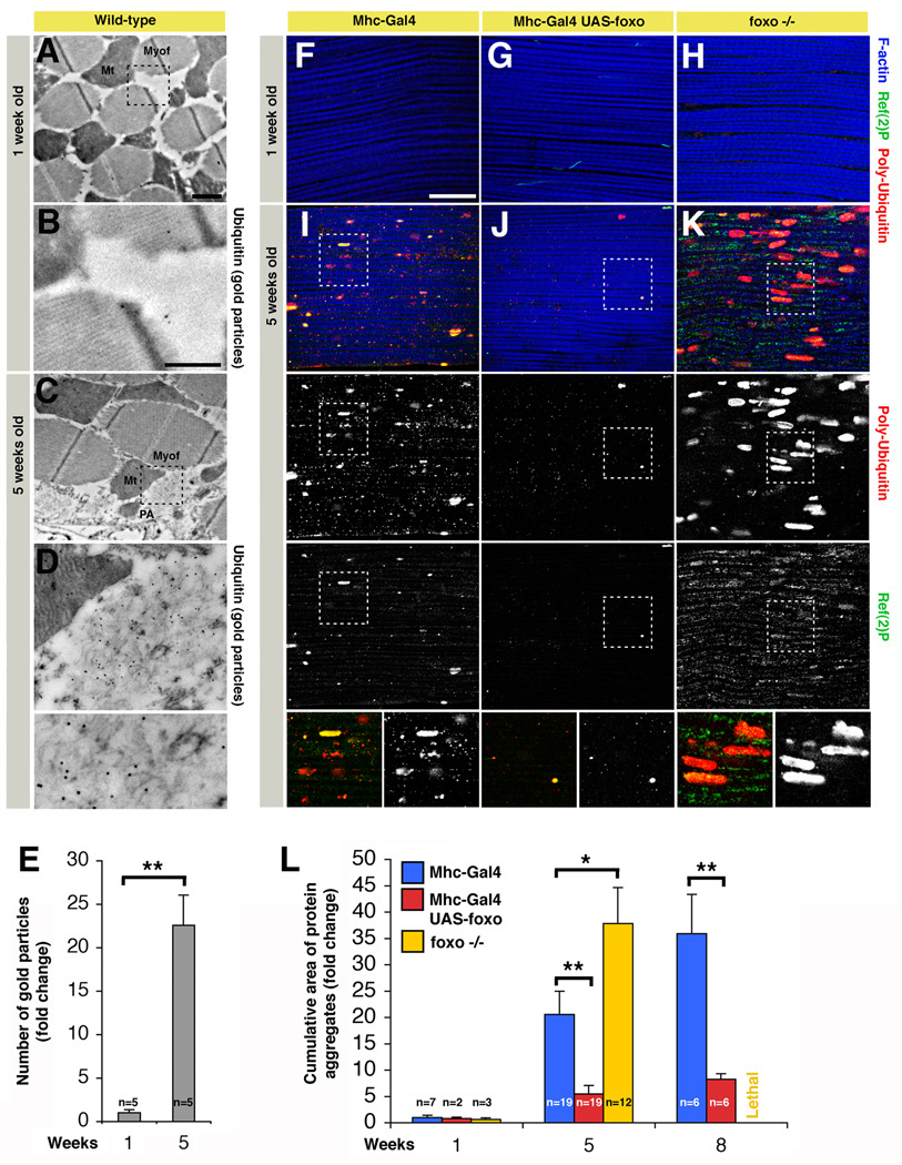
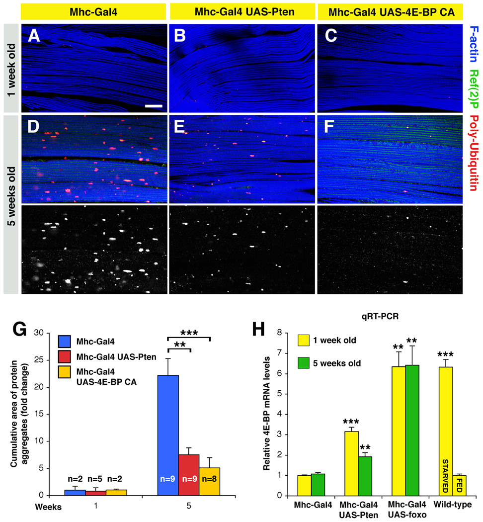
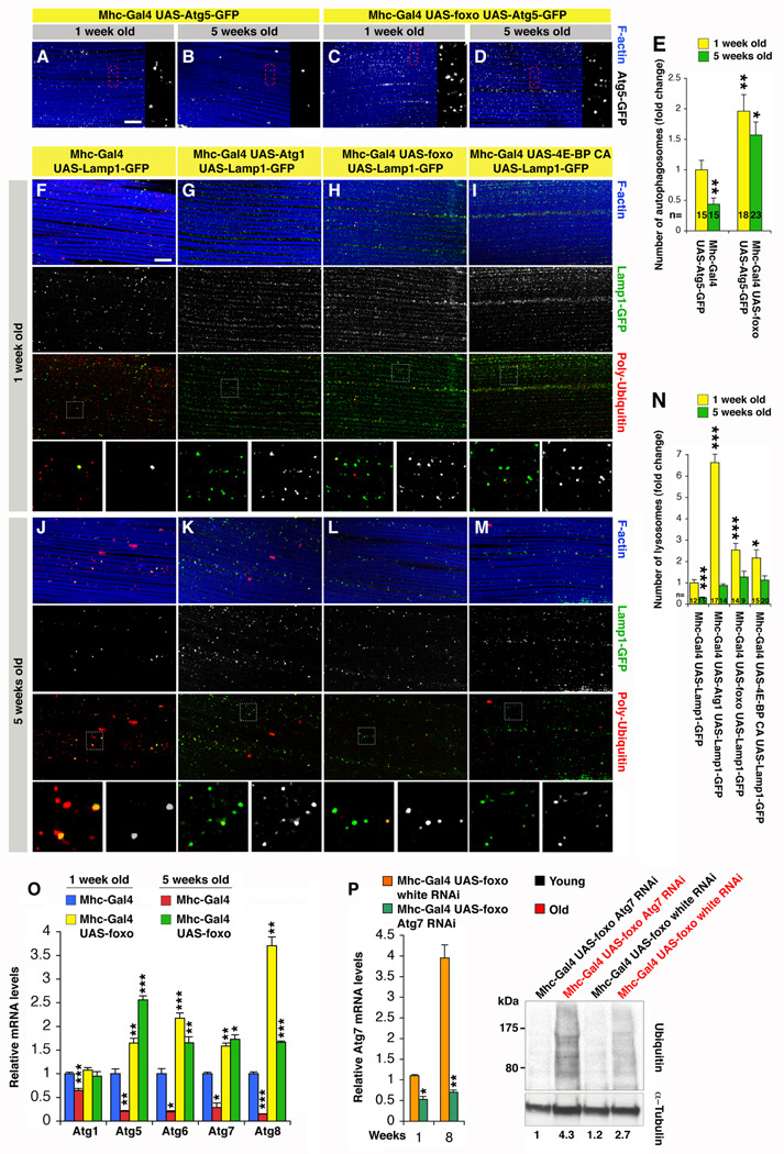
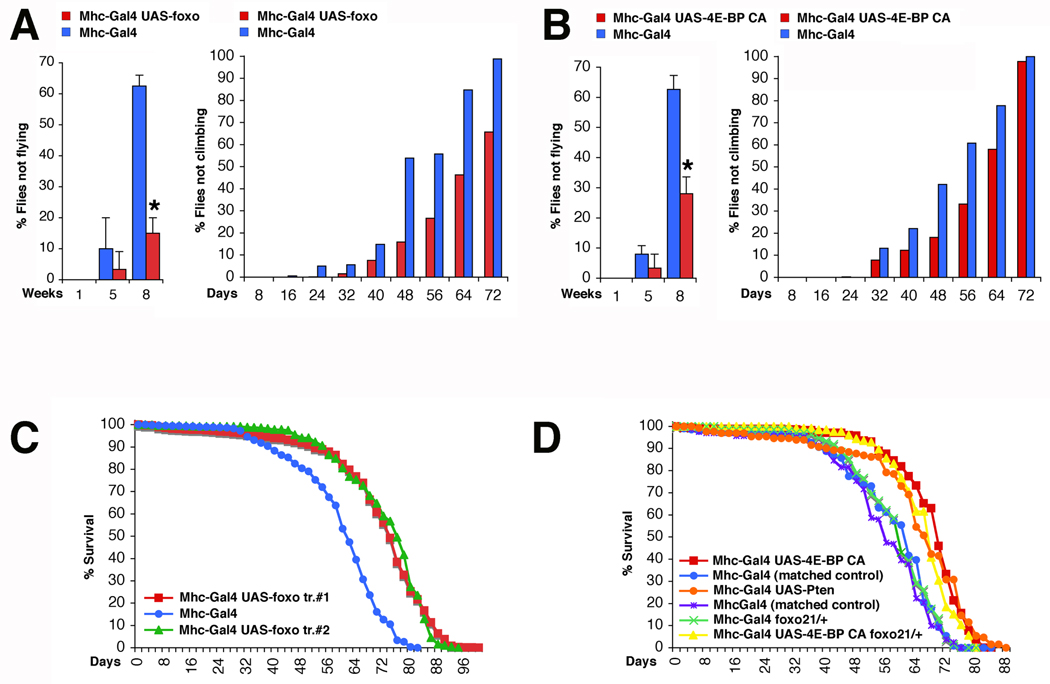
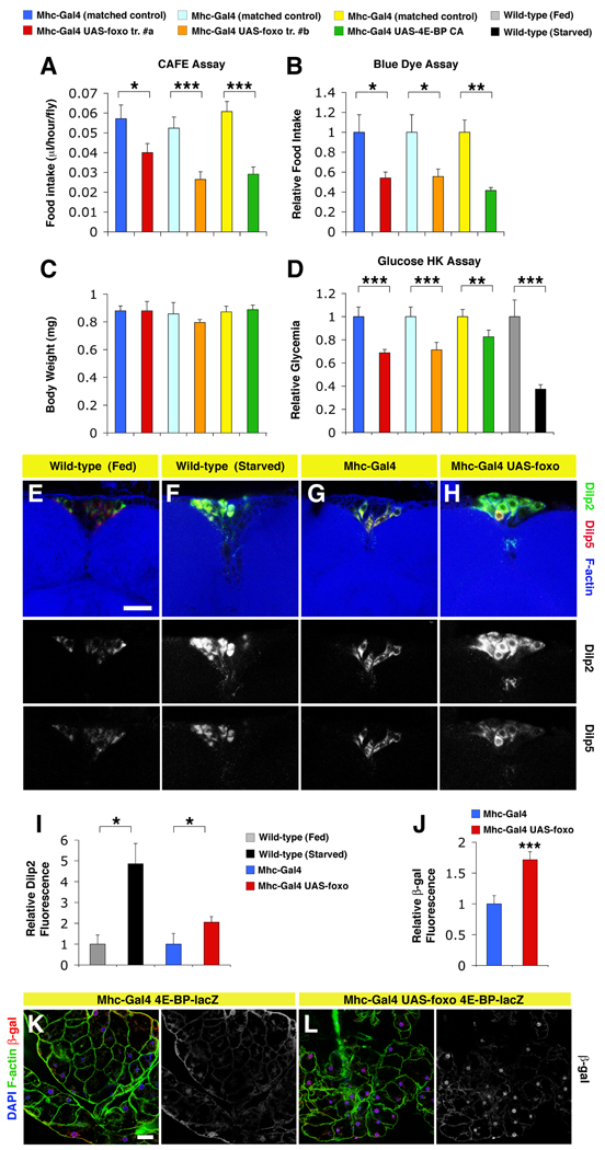
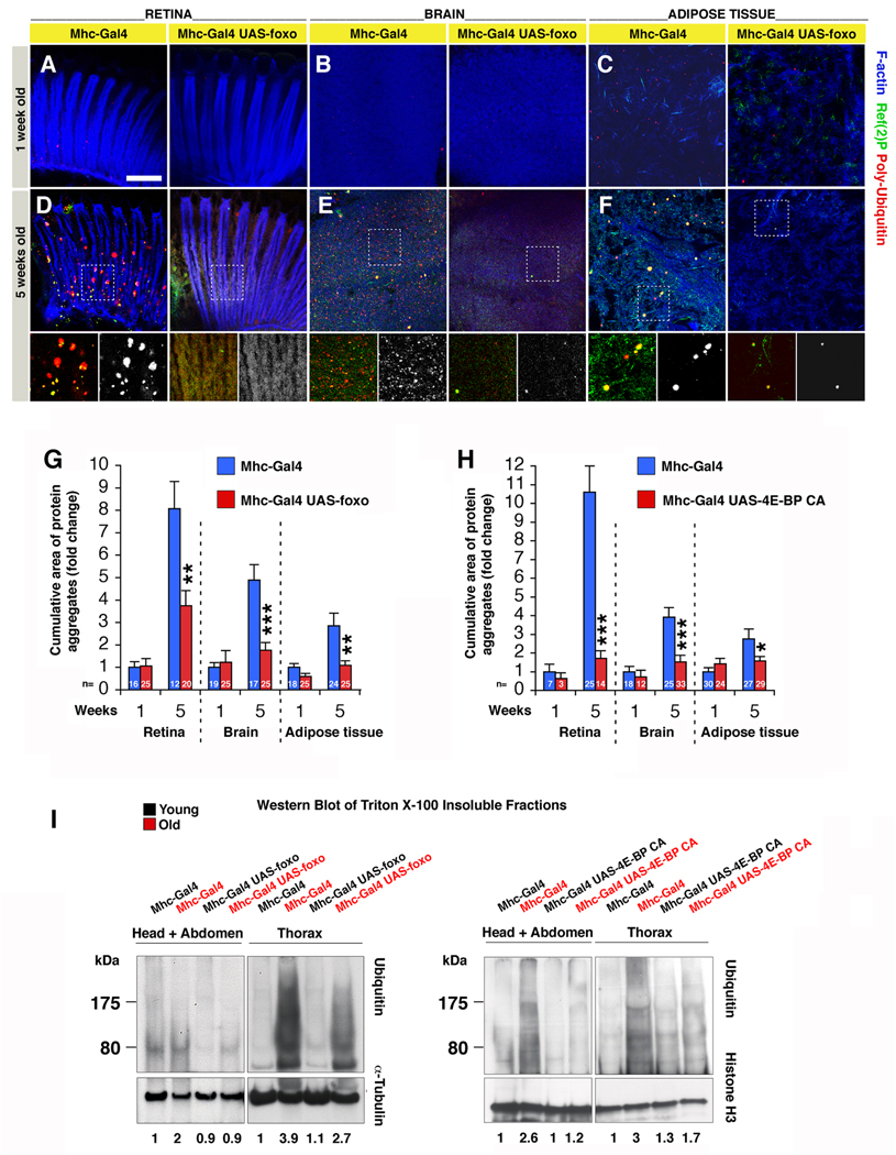
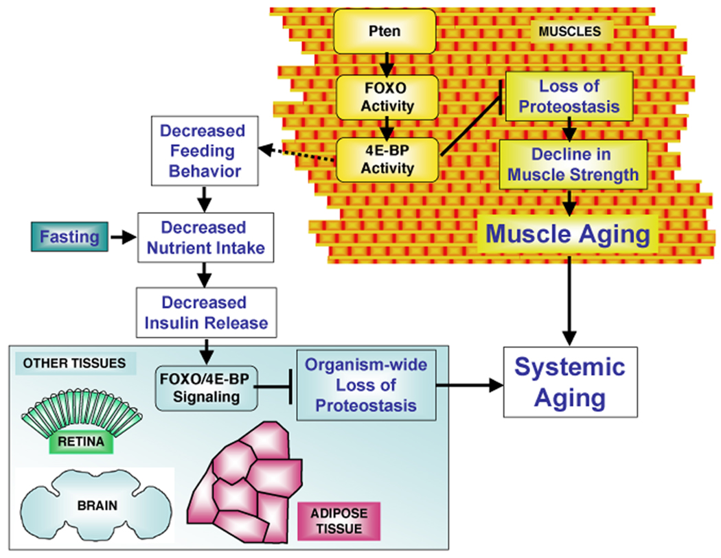
The progressive loss of muscle strength during aging is a common degenerative event of unclear pathogenesis. Although muscle functional decline precedes age-related changes in other tissues, its contribution to systemic aging is unknown. Here, we show that muscle aging is characterized in Drosophila by the progressive accumulation of protein aggregates that associate with impaired muscle function. The transcription factor FOXO and its target 4E-BP remove damaged proteins at least in part via the autophagy/lysosome system, whereas foxo mutants have dysfunctional proteostasis. Both FOXO and 4E-BP delay muscle functional decay and extend life span. Moreover, FOXO/4E-BP signaling in muscles decreases feeding behavior and the release of insulin from producing cells, which in turn delays the age-related accumulation of protein aggregates in other tissues. These findings reveal an organism-wide regulation of proteostasis in response to muscle aging and a key role of FOXO/4E-BP signaling in the coordination of organismal and tissue aging.


Figures

Figure 1
Figure 1. FOXO Signaling in Skeletal Muscles Preserves Proteostasis during Aging
(A–D) Electron micrographs of immunogold-labeled Drosophila skeletal muscles of wild-type flies at 1 (A–B) and 5 weeks of age (C–D). Protein aggregates (PA) are detected in the cytoplasm in proximity to mitochondria (Mt) and myofibrils (Myof) in old (C–D) but not young flies (A–B). Numerous gold particles (indicative of anti-Ubiquitin immunoreactivity) localize to filamentous structures at 5 weeks of age (C, D), while only few are present in muscles from young flies. Scale bars are 1 µm (A, C) and 500 nm (B, D). (E) The number of gold particles, indicative of Ubiquitin immunoreactivity, significantly increases in old age (sem is indicated with n; ** p<0.01). (F–K) Immunostaining of indirect flight muscles from flies with (UAS-foxo/+;Mhc-Gal4/+) or without (Mhc-Gal4/+) foxo overexpression at 1 week (F, G) and 5 weeks of age (I, J), and foxo homozygous null (MhcGal4, foxo21/25) flies (H, K). Poly-Ubiquitin (red) and p62/Ref(2)P (green) immunoreactivities reveal an increased deposition of aggregates containing poly-Ubiquitin proteins during aging in muscles of control flies (F, I), and to a lesser extent in muscles overexpressing foxo (G, J). Conversely, muscles from foxo null animals display an accelerated deposition of protein aggregates (H, K), in comparison with controls (F, I). Note the significant increase in the cumulative area of protein aggregates (indicative of both aggregate size and number) in (K) versus (I), and (I) versus (J), indicating that the control of protein homeostasis is linked to FOXO activity in muscles (quantification in L, sem is indicated with n; * p<0.05; ** p<0.01). Representative poly-Ubiquitin and Ref(2)P immunoreactivities are shown in insets. Phalloidin staining (blue) outlines F-actin, which is a component of muscle myofibrils. Scale bar is 20 μm (F-K). See also Figures S1, S2, and S3.
Figure 2
Figure 2. 4E-BP Preserves Proteostasis in Response to Pten/FOXO Signaling
(A–F) Immunostaining of muscles overexpressing Pten and constitutive active (CA) 4E-BP. In both cases, a decrease in the accumulation of Poly-Ubiquitin protein aggregates is observed at 5 weeks of age in comparison with age-matched controls, suggesting that these interventions can preserve proteostasis in aging muscles. Scale bar is 20 µm. Hsp70 overexpression has instead limited effects (Figure S4, Tables S1 and S2). (G) A reduction in the cumulative area of protein aggregates is observed upon increased activity of either Pten or 4E-BP in comparison with controls (sem is indicated with n; ** p<0.01; *** p<0.001). (H) Relative quantification of Thor/4E-BP mRNA levels from thoraces of syngenic flies at 1 and 5 weeks of age. A significant increase in 4E-BP expression is detected in response to fasting, and Pten and FOXO activity (**p<0.01; ***p<0.001; sem is indicated with n=4).
Figure 3
Figure 3. FOXO and 4E-BP Regulate Proteostasis at Least in Part via the Autophagy/ Lysosome System
(A–E) Immunostaining of muscles expressing the marker of autophagosomes Atg5-GFP reveals a significant increase in their number (E) and maintenance at 1 and 5 weeks of age upon foxo overexpression (C, D), in comparison with controls (A, B). In (E), sem is indicated with n; *p<0.05 and **p<0.01. (F–N) Immunostaining of muscles expressing the lysosomal marker Lamp1-GFP, and overexpressing either Atg1, foxo, and 4E-BP CA. Note an increase in the number of lysosomes (N) at both 1 (G–I) and 5 weeks of age (K–M), which inversely correlates with poly-Ubiquitin immunoreactivity, in comparison with control muscles (F, J). Scale bar is 10 µm (A–D and F–M). In (N), sem is indicated with n; *p<0.05 and ***p<0.001. (O) Relative mRNA levels of autophagy genes from thoraces of 1 and 5 weeks old flies decrease during normal muscle aging while their expression increases and persists in response to FOXO. Sem is indicated with n=4; *p,0.05, **p<0.01 and ***p<0.001. (P) RNAi treatment against Atg7 results in a ~50% knock-down of its mRNA levels in muscles and partially impairs FOXO-mediated proteostasis, as indicated by the increased detection of Ubiquitin-conjugated proteins in Triton X-100 insoluble fractions at 8 weeks (old, red) in comparison with mock treated (white RNAi) and young flies (1 week old, black). Normalized values based on α-Tubulin levels are indicated.
Figure 4
Figure 4. FOXO/4E-BP Signaling Preserves Muscle Function and Extends Lifespan
(A) Muscle function gradually decreases during aging as indicated by an increase in the percentage of flies with climbing and flight defects. However, foxo preserves their function in comparison with controls (Flight ability: n[flies]= 10 (week 1 and 5) and 30 (week 8) with n[batch]= 3 (week 1 and 5) and 2 (week 8); sd is indicated and *p<0.05. Climbing ability: (n[Mhc-Gal4/+]=1264, n[Mhc-Gal4/UAS-foxo]=966, with n indicating the number of flies at day 1; p<0.001). (B) Similar to FOXO, 4E-BP activity also results in decreased age-related flight and climbing deficits in comparison with controls (Flight ability: n[flies]≥10 (week 1 and 5) and 25 (week 8) with n[batch]≥3 (week 1 and 5) and 2 (week 8); sd is indicated and *p<0.05. Climbing ability: (n[Mhc-Gal4/+]=204, n[Mhc-Gal4/UAS-4E-BP CA]=403, p<0.001). (C) Survival of flies during aging. Foxo overexpression in muscles significantly extends the median and maximum lifespan (Median and maximum lifespan: Mhc-Gal4/+=~61 and 82 days (n=1264); UAS-foxo tr.#1/+;Mhc-Gal4/+=~73 and 100 days (n=1184); Mhc-Gal4/UAS-foxo tr.#2=~76 and 94 days (n=966); p<0.001). (D) Lifespan of flies with increased Pten and 4E-BP activity in muscles is extended in comparison with matched controls (Median and maximum lifespan of 4E-BP: Mhc-Gal4/+=~63 and 78 days (n=204); Mhc-Gal4/UAS-4E-BP CA =~71 and 84 days (n=403); Pten: Mhc-Gal4/+=~55 and 76 days (n=162); Mhc-Gal4/UAS-Pten=~66 and 88 days (n=130); p<0.001). Similar increase in lifespan is brought about by 4E-BP CA overexpression in foxo21 heterozygous null flies. See also Figures S5 and S7.
Figure 5
Figure 5. FOXO Signaling in Muscles Partially Mimics Systemic Metabolic Changes Associated with Fasting by Modulating Feeding Behavior
(A–C) Flies in which FOXO/4E-BP activity has been altered specifically in muscles consume less food than matched controls. Food consumption was determined via capillary feeding CAFÉ assay over 2 hours periods (A), and by monitoring the ingestion of blue colored food in 24 hours (B). Error bars represent sem with n[measurements]=44, 46, 52, 37, 103, and 61 in (A) and n=2 in (B), with * p<0.05; ** p<0.01; *** p<0.001. Decreased feeding does not result from developmental defects, as indicated by similar body weights of flies analyzed (C; error bars represent sd with n≥3). (D) Relative glucose levels (glycemia) in the hemolymph of flies overexpressing either foxo or 4E-BP CA in muscles, and matched controls. Manipulation of FOXO/4E-BP signaling in muscles brings about a reduction of glycemia similar in part to that of wild-type flies starved for 24 hours, as estimated with the Glucose Hexokinase Assay (sem is indicated with n=5, and ** p<0.01; *** p<0.001). (E–H) Immunostaining of Dilp-producing median neurosecretory cells in the brain of starved wild-type flies, flies overexpressing foxo in muscles, and controls. Increase in the immunoreactivity of Insulin-like peptides Dilp2 (green) is detected in producing cells in response to either starvation (F) or foxo overexpression in muscles (H), in comparison respectively with fed wild-type flies (E), and controls with no foxo overexpression in muscles (G). Smaller changes in Dilp5 levels are observed. Phalloidin staining (blue) detects F-actin (scale bar is 20 µm; images in E–H have the same magnification). (I) Quantification of the intensity of staining indicates that differences in Dilp2 fluorescence are significant (sd is indicated with n[measurements]=35, 69, 37, and 96 from n[brains]=2, 4, 3, and 4; *p<0.05). (J–L) Quantification and immunostaining of adipose tissue (peripheral fat body of the abdomen) from 2 weeks old flies. (J) Note a significant increase in nuclear β-galactosidase immunoreactivity (red) in the adipose tissue from flies with a nuclear 4E-BP-lacZ reporter and foxo overexpression in muscles (L), in comparison with controls (K). F-actin (green) and DAPI staining (indicative of nuclei, blue) are shown. Scale bar is 20 µm. In (J), sem is indicated with n=20 and *** p<0.001.
Figure 6
Figure 6. Systemic Proteostasis is Remotely Controlled by FOXO/4E-BP Signaling in Muscles
(A–F) Aggregates of poly-Ubiquitin proteins accumulate during aging in the retina (A, D), brain (B, E), and the adipose tissue (C, F) of control flies (Mhc-Gal4/+), but to a lesser extent in tissues from flies overexpressing foxo in muscles (UAS-foxo/+;Mhc-Gal4/+), as indicated by poly-Ubiquitin (red) and p62/Ref(2)P (green) stainings. Phalloidin staining (blue) outlines F-actin. Note that Mhc-Gal4 does not drive transgene expression in these tissues (Figure S1). Scale bar is 10 μm. (G–H) The age-related increase in the cumulative area of protein aggregates is significantly less prominent in tissues from flies overexpressing foxo (G) or 4E-BP CA (H) in muscles in comparison with controls (sem is indicated with n; *p<0.05. **p<0.01, and ***p<0.001). (I) Ubiquitin levels (indicative of protein aggregates) are detected in Triton X-100 insoluble fractions from thoraces, and head and abdominal tissues from flies overexpressing foxo in muscles or control flies at 1 (young, black) and 8 (old, red) weeks of age. Ubiquitin levels are increased in old flies in comparison with young flies in extracts from both muscles (thoraces) and non-muscle tissues (heads and abdomens). However, flies overexpressing foxo in muscles have reduced deposition of protein aggregates at 8 weeks of age in both muscles and non-muscle tissues. Similar results are obtained in response to increased 4E-BP activity in muscles (I), but not Hsp70 (Figure S5). Quantification of Ubiquitin-conjugated proteins normalized to α-Tubulin or Histone H3 levels is indicated. See also Figures S1, S5 and S6.
Figure 7
Figure 7. FOXO/4E-BP Signaling in Muscles Controls Proteostasis and Systemic Aging
Muscle aging is characterized by protein damage and accumulation of cytoplasmic aggregates. Loss of protein homeostasis (proteostasis) associates with the progressive decrease in muscle strength and can affect the lifespan of the organism. Pten/FOXO signaling induces multiple targets, including several folding chaperones and the regulator of protein translation 4E-BP. FOXO/4E-BP activity regulates muscle proteostasis at least in part via the autophagy/lysosome pathway of protein degradation, preserves muscle function, and extends lifespan. In addition, FOXO/4E-BP signaling in muscles decreases feeding behavior that, similar to fasting, results in reduced Insulin release from producing cells. This in turn promotes FOXO and 4E-BP activity in other tissues, preserving proteostasis organism-wide and mitigating systemic aging.

References

    1. Arndt V, Dick N, Tawo R, Dreiseidler M, Wenzel D, Hesse M, Furst DO, Saftig P, Saint R, Fleischmann BK, et al. Chaperone-Assisted Selective Autophagy Is Essential for Muscle Maintenance. Curr Biol. 2010;20:143–148. - PubMed
    1. Augustin H, Partridge L. Invertebrate Models of Age-Related Muscle Degeneration. Biochim Biophys Acta. 2009;1790:1084–1094. - PubMed
    1. Bodine SC, Stitt TN, Gonzalez M, Kline WO, Stover GL, Bauerlein R, Zlotchenko E, Scrimgeour A, Lawrence JC, Glass DJ, Yancopoulos GD. Akt/mTOR pathway is a crucial regulator of skeletal muscle hypertrophy and can prevent muscle atrophy in vivo. Nat Cell Biol. 2001;3:1014–1019. - PubMed
    1. Boyle PA, Buchman AS, Wilson RS, Leurgans SE, Bennett DA. Association of muscle strength with the risk of Alzheimer disease and the rate of cognitive decline in community-dwelling older persons. Arch Neurol. 2009;66:1339–1344. - PMC - PubMed
    1. Broughton SJ, Slack C, Alic N, Metaxakis A, Bass TM, Driege Y, Partridge L. DILP-producing median neurosecretory cells in the Drosophila brain mediate the response of lifespan to nutrition. Aging Cell. 2010;9:336–346. - PMC - PubMed

Publication types

Substances

LinkOut - more resources