Comparative Study
. 2012 Apr 19;156(2):133-8.
doi: 10.1016/j.ijcard.2010.10.025. Epub 2010 Dec 15.

The temporal and spatial expression patterns of ABCG2 in the developing human heart

Affiliations
Comparative Study

The temporal and spatial expression patterns of ABCG2 in the developing human heart

Marah Alfakir et al. Int J Cardiol. .

Abstract

Background: The discovery that the adult heart is not a terminally differentiated organ and contains stem/progenitor cells has important implications for the development of cellular therapeutics to treat heart disease. Moreover the discovery of cardiac stem cells might be important in furthering our understanding of both normal and abnormal cardiac development and yet little is known about these cell populations in the developing human heart, which we have focused on in this study.

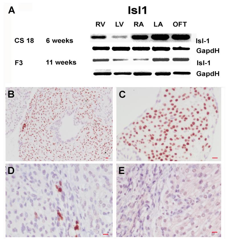
Methods: The presence of ABCG2 and islet-1 expressing cells in human heart was determined using immunohistochemistry and RT-PCR (and western blotting for ABCG2). Cardiac SP cells were isolated using FACS. Co-localisation immunohistochemistry was used to determine if ABCG2 positive cells expressed other known stem/progenitor cell, endothelial markers or cardiac markers.

Results: We observed that ABCG2 expressing cells show a difference in both their temporal and spatial patterns of expression from Islet-1 expressing cardiac progenitors. We identified rare cells that expressed both ABCG2 and markers of other cell lineages including CD31, CD34 and alpha-actinin. We also noted the presence of cells that only expressed ABCG2. We isolated cardiac SP cells and confirmed the SP cell phenotype.

Conclusions: Our results suggest that the developing human heart contains at least two distinct cardiac stem/progenitor cell populations one of which, the ABCG2 positive cells, can be readily isolated, suggesting that this tissue could be a useful source of cardiac stem cells.


Figures

Figure 1
Figure 1
ABCG2 is expressed in the developing human heart. (A) Representative RT-PCR analysis for ABCG2 expression from CS18 (6 weeks pc) through to F5 (13 weeks pc) comparing the expression of ABCG2 mRNA in distinct anatomical cardiac regions. Note at CS18 the expression appears to be similar in all cardiac regions with the exception of the OFT where it is slightly elevated. At CS23 (8 weeks pc) expression is down regulated in the LV compared with the other anatomical regions and in the fetal stages the expression of ABCG2 is reduced in LV, LA compared with the RV, RA and OFT. GapdH was used as a loading control. (B) FACS profile of CSP cells from an F1 heart (9 weeks pc). (C) Inhibition of the SP cell phenotype after addition of FTC. Abbreviations: pc: post conception; SP, side population: RV, right ventricle; LV, left ventricle; RA, right atria; LA, left atria; OFT, outflow tract; FTC, fumitremorgin C; RT-PCR, reverse transcriptase polymerase chain reaction. IHC analysis for ABCG2, ABCG2 expressing cells are located throughout the human embryonic and fetal heart. Shown are representative images taken of cardiac tissue stained with an anti-ABCG2 antisera. (D and F) ABCG2 expressing cells in the embryonic heart at CS23 (8 weeks pc) (D) and in the ventricles at F1 (9 weeks pc) shown here in the LV (F). Note the absence of staining in the no primary negative control (E). ABCG2 was visualised with 3’-diaminobenzidine (brown), all sections were counterstained with haematoxylin (blue). Magnifications: size bar = 10 μm for all images; Abbreviations: pc: post conception; SP, side population: RV, right ventricle; LV, left ventricle; RA, right atria; LA, left atria; OFT, outflow tract; FTC, fumitremorgin C; RT-PCR, reverse transcriptase polymerase chain reaction, IHC, immunohistochemical.
Figure 2
Figure 2
Islet-1 is expressed during development of the human heart. (A) RT-PCR results for islet1 mRNA expression at CS18 (6 weeks pc) and F3 (11 weeks pc). Immunohistochemical analysis for islet-1 expressing cells, large numbers of isl+ cells are present in the OFT (B) and RA (C) at CS23 (8 weeks pc). Small numbers of isl+ cells are also present in ventricles at F6 (14 weeks pc) (D). Note the absence of staining in the no primary control, F6 (14 weeks pc) ventricle (E). Islet1 was visualised with 3’-diaminobenzidine (brown) and sections were counterstained with haematoxylin (blue) Magnifications size bar = 10μm for all images. Abbreviations: RT-PCR: reverse transcription–polymerase chain reaction, pc: post conception, Isl+: islet-1 positive, RA: right atria, OFT: outflow tract.
Figure 3
Figure 3
Representative results for co-expression analysis for ABCG2 and other stem, progenitor endothelial and cardiac markers. No cells expressed islet −1 and ABCG2 in the OFT (A) or RA (B) at CS23 (8 weeks pc). Rare cells that expressed both CD31 and ABCG2 are expressed in the embryonic heart at CS23 (8 weeks pc) in the RA, note a cell expressing ABCG2 alone and a dual labelled cell expressing both ABCG2 and CD31 (C). Small numbers of cells that express ABCG2 and CD34 are present in the developing heart, in the LV at CS23 (8 weeks pc) (D). Rare cells express both ABCG2 and alpha-actinin in the fetal heart at F1 (9weeks pc) (E). Images A,B.C, and E, ABCG2 visualised with 3’-diamionbenzidine (brown), islet-1, CD31 and alpha-actinin (sarcomeric) visualised with Vector blue (blue). Image D, ABCG2 visualised with Vector NovaRED (red), CD34 visualised with Vector SG blue/grey (grey). Magnifications: Size bar = 10μm Abbreviations: OFT: outflow tract, LV: left ventricle, pc: post conception, RA: right atria, LA: left atria, RV: right ventricle.

References

    1. Beltrami A, Barlucchi L, Torella D, et al. Adult cardiac stem cells are multipotent and support myocardial regeneration. Cell. 2003;114:763–776. - PubMed
    1. Oh H, Bradfute SB, Gallardo TD, et al. Cardiac progenitor cells from adult myocardium: homing, differentiation, and fusion after infarction. PNAS. 2003;100:12313–12318. - PMC - PubMed
    1. Laugwitz KL, Moretti A, Lam J, et al. Postnatal isl1+ cardioblasts enter fully differentiated cardiomyocyte lineages. Nature. 2005;433:647–653. - PMC - PubMed
    1. Messina E, De Angelis L, Frati G, et al. Isolation and expansion of adult cardiac stem cells from human and murine heart. Circ Res. 2004;95:911–921. - PubMed
    1. Martin CM, Meeson AP, Roberston SM, et al. Persistent expression of the ATP-binding cassette transporter, Abcg2, Identifies cardiac SP cells in the developing and adult heart. Dev Biol. 2004;265:262–275. - PubMed

Publication types

MeSH terms

Substances