. 2011 Feb 11;406(1):75-91.
doi: 10.1016/j.jmb.2010.11.031. Epub 2010 Nov 25.

Substrate-induced conformational changes occur in all cleaved forms of caspase-6

Affiliations

Substrate-induced conformational changes occur in all cleaved forms of caspase-6

Sravanti Vaidya et al. J Mol Biol. .

Abstract

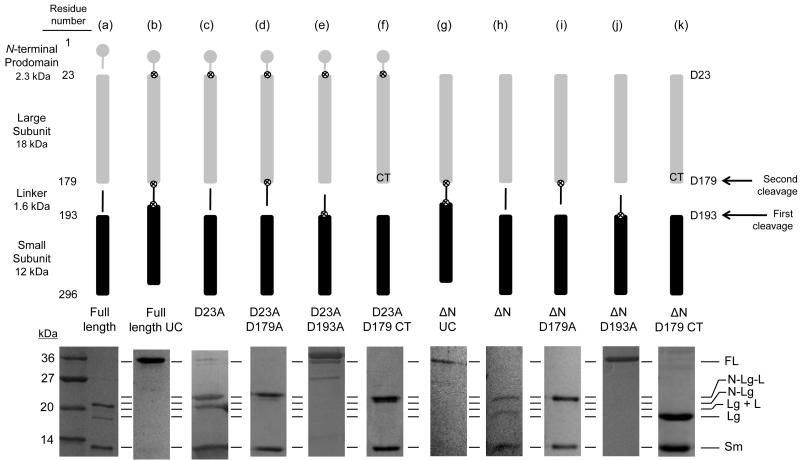
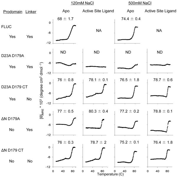
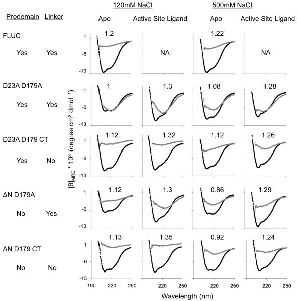
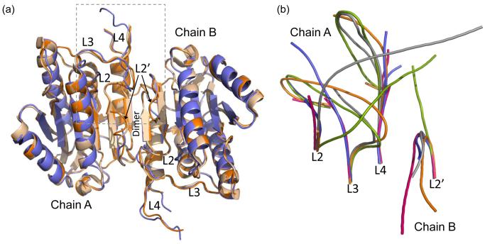
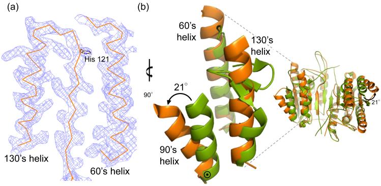
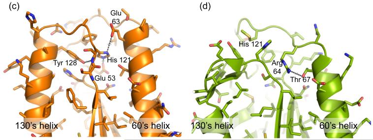
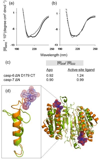
Caspase-6 is an apoptotic cysteine protease that also governs disease progression in Huntington's and Alzheimer's diseases. Caspase-6 is of great interest as a target for treatment of these neurodegenerative diseases; however, the molecular basis of caspase-6 function and regulation remains poorly understood. In the recently reported structure of caspase-6, the 60's and 130's helices at the base of the substrate-binding groove extend upward, in a conformation entirely different from that of any other caspase. Presently, the central question about caspase-6 structure and function is whether the extended conformation is the catalytically competent conformation or whether the extended helices must undergo a large conformational rearrangement in order to bind substrate. We have generated a series of caspase-6 cleavage variants, including a novel constitutively two-chain form, and determined crystal structures of caspase-6 with and without the intersubunit linker. This series allows evaluation of the role of the prodomain and intersubunit linker on caspase-6 structure and function before and after substrate binding. Caspase-6 is inherently more stable than closely related caspases. Cleaved caspase-6 with both the prodomain and the linker present is the most stable, indicating that these two regions act in concert to increase stability, but maintain the extended conformation in the unliganded state. Moreover, these data suggest that caspase-6 undergoes a significant conformational change upon substrate binding, adopting a structure that is more like canonical caspases.


Figures

Fig. 1
Fig. 1
Caspase-6 cleavage site-blocking variants. Cartoon representation of expression constructs showing cleavable (gap) or blocked ⊗ caspase cleavage sites (upper panel). Symbols for protein domains include prodomain (gray circle), large subunit (gray bar), intersubunit linker (black line), and small subunit (black bar). Aspartic acid residues mutated to alanine are depicted as ⊗. Residue numbers for each domain are indicated at the left of the upper panel. SDS-PAGE analysis of expressed constructs matured in E. coli cells and the observed cleavage patterns (lower panel). (a) Full-length caspase-6. (b) Full-length uncleavable caspase-6 variant (FLUC) D23A/D179A/D193A completely blocks zymogen processing. (c) D23A. (d) D23A/D179A. (e) D23A/D193A. (f) D23A/D179 Constitutive Two-chain (CT) variant expresses the prodomain-large subunit protein inclusive of residue 179 independently of the small subunit, which is expressed from an introduced ribosome-binding site and start codon at residue 193. (g-k) Expression constructs similar to b-f but lack the coding sequence for the N-terminal prodomain. FL= Full Length; N = N-terminal prodomain; Lg = Large subunit; Sm = Small subunit, L = Intersubunit linker. The order of cleavage events leading to activation is indicated by arrows on the right.
Fig. 2
Fig. 2
Circular dichroism (CD) of caspase-6 cleavage variants. (a) Thermal denaturation profiles of caspase-6 cleavage variants unliganded (apo) or with the active-site ligand VEID in physiological (120 mM) or high (500 mM) NaCl concentrations. The measured Tm did not vary between runs, however run-to-run variation in the shape and slope of the pre-melt transition precluded meaningful interpretation of this region of the melting curve. (b) CD spectra of caspase-6 cleavage variants at 12°C (black) or 90°C (gray). The [θ]208/[θ]222 values at 12°C are indicated above each spectra. Each variant was measured using two independently prepared and concentrated samples on two different days. The spectral features were observed in duplicate spectra and a representative spectrum for each variant is shown. NA Not applicable due to inability to functionally verify binding of VEID to the active site. ND Not Detectable.
Fig. 2
Fig. 2
Circular dichroism (CD) of caspase-6 cleavage variants. (a) Thermal denaturation profiles of caspase-6 cleavage variants unliganded (apo) or with the active-site ligand VEID in physiological (120 mM) or high (500 mM) NaCl concentrations. The measured Tm did not vary between runs, however run-to-run variation in the shape and slope of the pre-melt transition precluded meaningful interpretation of this region of the melting curve. (b) CD spectra of caspase-6 cleavage variants at 12°C (black) or 90°C (gray). The [θ]208/[θ]222 values at 12°C are indicated above each spectra. Each variant was measured using two independently prepared and concentrated samples on two different days. The spectral features were observed in duplicate spectra and a representative spectrum for each variant is shown. NA Not applicable due to inability to functionally verify binding of VEID to the active site. ND Not Detectable.
Fig. 3
Fig. 3
Structures of apo-caspase-6 ΔN D179A and ΔN D179 CT. (a) Superposition of caspase-6 ΔN D179A (orange) and ΔN D179 CT (tan) and an independently determined caspase-6 structure (2WDP, blue). The boxed region indicates the substrate binding groove region that is shown in (b). (b) Superposition of unliganded mature caspase-6 ΔN D179A (orange) with liganded mature caspase-7 (1F1J, green) allosterically inhibited caspase-7 (1SHJ, pink), caspase-7 zymogen (1GQF, blue), and unliganded caspase-7 (1K86, gray) focusing on the substrate-binding loop region including loops L2, L3 and L4 from chain A of the dimer and L2’ from chain B of the dimer. Disordered regions not observed in the crystal structure account for the discontinuity in the middle of loops L3 and L4 in some of the structures.
Fig. 4
Fig. 4
Unique structural features of caspase-6. (a) 2Fo-Fc electron density map contoured at 1 σ in the region of the 60’sand 130’s helices. Cα trace is shown in orange. (b) Comparison of mature ligand-free caspase-6 60’s, 90’s and 130’s helices (orange) to the homologous region of mature ligand-free caspase-7 (1K86, green). ⊙ denotes the hinge around which the 90’s helix pivots by 21°. (c) Interactions (dashes) holding the 60’s and 130’s network of helices together are mediated by the inactive conformation of catalytic-dyad residue, His 121. (d) The homologous region to (c) in the active-site liganded caspase-7 structure with caspase-6 numbering shown. Indicated residues (drawn in sticks) have the same amino acid identity in both caspase-6 and caspase-7, although the numbering is different. R64, T67 and H121 in caspase-6 numbering are R87, T90 and H144 in caspase-7 numbering. (e) Sequence alignment for the 60’s, 90’s and 130’s helices for all caspases. Amino acid numbering for each caspase is indicated. Strictly conserved residues (orange letters), important network residues (gray highlight) and the catalytic histidine (yellow highlight) in apoptotic initiators, executioners (Exec) and inflammatory (Inflam) caspases are shown. (f) Surface of caspase-6 near the 90’s helix (drawn with 2WDP coordinates, in which the 90’s helix side chains are better resolved) showing the pocket between the 90’s and 130’s helices
Fig. 4
Fig. 4
Unique structural features of caspase-6. (a) 2Fo-Fc electron density map contoured at 1 σ in the region of the 60’sand 130’s helices. Cα trace is shown in orange. (b) Comparison of mature ligand-free caspase-6 60’s, 90’s and 130’s helices (orange) to the homologous region of mature ligand-free caspase-7 (1K86, green). ⊙ denotes the hinge around which the 90’s helix pivots by 21°. (c) Interactions (dashes) holding the 60’s and 130’s network of helices together are mediated by the inactive conformation of catalytic-dyad residue, His 121. (d) The homologous region to (c) in the active-site liganded caspase-7 structure with caspase-6 numbering shown. Indicated residues (drawn in sticks) have the same amino acid identity in both caspase-6 and caspase-7, although the numbering is different. R64, T67 and H121 in caspase-6 numbering are R87, T90 and H144 in caspase-7 numbering. (e) Sequence alignment for the 60’s, 90’s and 130’s helices for all caspases. Amino acid numbering for each caspase is indicated. Strictly conserved residues (orange letters), important network residues (gray highlight) and the catalytic histidine (yellow highlight) in apoptotic initiators, executioners (Exec) and inflammatory (Inflam) caspases are shown. (f) Surface of caspase-6 near the 90’s helix (drawn with 2WDP coordinates, in which the 90’s helix side chains are better resolved) showing the pocket between the 90’s and 130’s helices
Fig. 5
Fig. 5
Mutants to probe the unique 90’s helix conformation. (a) Models of the 90’s and 130’s helices from caspase-6 (orange) in the open conformation and caspase-7 (green) in closed conformation. Caspase-6 positions 96 and 139 are shown mutated from Leu to Trp. Caspase-6 L96 and L139 correspond to residues M116 and L162 respectively in caspase-7. L96 and L139 can accommodate at least one rotomer of Trp in the open conformation, but no rotomers of Trp in the closed conformation, as evidenced by the observed clashes between the introduced Trp and adjacent side chains. (b) The kinetic parameters for caspase-6 ΔN D179A (WT), ΔN L96W D179A (L96W) and ΔN L139W D179A (L139W).
Fig. 6
Fig. 6
Comparison of caspase-6 and caspase-7 structures and CD spectra in the presence or absence of the active-site ligands VEID or DEVD. (a) CD spectra of caspase-6 bound to active-site ligand VEID (gray) or in the apo state with no ligand bound (black). (b) CD spectra of caspase-7 bound to active-site ligand DEVD (gray) or in the apo state with no ligand bound (black). (c) Comparision of [θ]208/[θ]222 for apo and substrate-bound states of caspase-6 and caspase-7. (d) Superposition of mature ligand-free caspase-6 ΔN D179CT (orange) with caspase-7 (1F1J, green) bound to the substrate-like ligand DEVD (purple dots).

References

    1. Fernandes-Alnemri T, Litwack G, Alnemri E. Mch2, a new member of the apoptotic Ced-3/Ice cysteine protease gene family. Cancer research. 1995;55(13):2737. - PubMed
    1. Fuentes-Prior P, Salvesen GS. The protein structures that shape caspase activity, specificity, activation and inhibition. Biochem J. 2004;384(Pt 2):201–32. - PMC - PubMed
    1. Fujita E, et al. Caspase-9 processing by caspase-3 via a feedback amplification loop in vivo. Cell Death Differ. 2001;8(4):335–44. - PubMed
    1. Suzuki A, et al. Regulation of caspase-6 and FLIP by the AMPK family member ARK5. Oncogene. 2004;23(42):7067–75. - PubMed
    1. Takahashi A, et al. Cleavage of lamin A by Mch2 alpha but not CPP32: multiple interleukin 1 beta-converting enzyme-related proteases with distinct substrate recognition properties are active in apoptosis. Proc Natl Acad Sci U S A. 1996;93(16):8395–400. - PMC - PubMed

Publication types

LinkOut - more resources