

PET/CT Imaging and Radioimmunotherapy of Prostate Cancer
- PMID: 21111858
- PMCID: PMC3392994
- DOI: 10.1053/j.semnuclmed.2010.08.005
PET/CT Imaging and Radioimmunotherapy of Prostate Cancer
Abstract
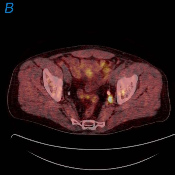
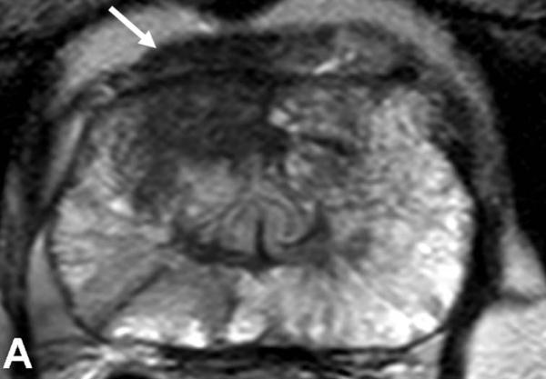
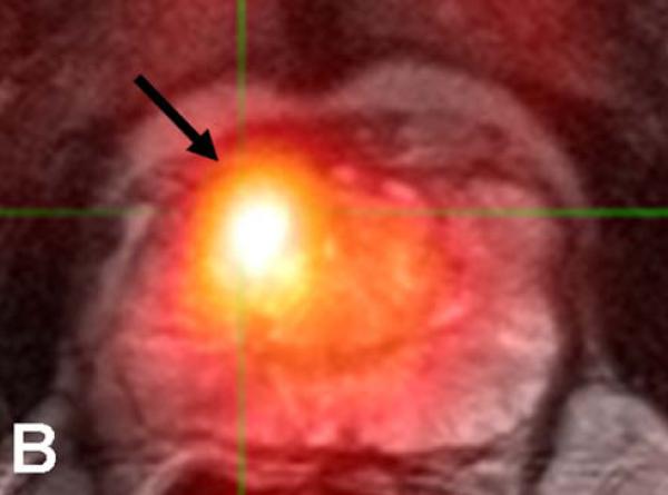
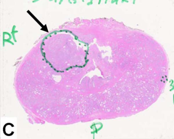
Prostate cancer is a common cancer in men and continues to be a major health problem. Imaging plays an important role in the clinical management of patients with prostate cancer. An important goal for prostate cancer imaging is more accurate disease characterization through the synthesis of anatomic, functional, and molecular imaging information. Positron emission tomography (PET)/computed tomography (CT) in oncology is emerging as an important imaging tool. The most common radiotracer for PET/CT in oncology, (18)F-fluorodeoxyglucose (FDG), is not very useful in the imaging of prostate cancer. However, in recent years other PET tracers have improved the accuracy of PET/CT imaging of prostate cancer. Among these, choline labeled with (18)F or (11)C, (11)C-acetate, and (18)F-fluoride has demonstrated promising results, and other new radiopharmaceuticals are under development and evaluation in preclinical and clinical studies. Large prospective clinical PET/CT trials are needed to establish the role of PET/CT in prostate cancer patients. Because there are only limited available therapeutic options for patients with advanced metastatic prostate cancer, there is an urgent need for the development of more effective treatment modalities that could improve outcome. Prostate cancer represents an attractive target for radioimmunotherapy (RIT) for several reasons, including pattern of metastatic spread (lymph nodes and bone marrow, sites with good access to circulating antibodies) and small volume disease (ideal for antigen access and antibody delivery). Furthermore, prostate cancer is also radiation sensitive. Prostate-specific membrane antigen is expressed by virtually all prostate cancers, and represents an attractive target for RIT. Antiprostate-specific membrane antigen RIT demonstrates antitumor activity and is well tolerated. Clinical trials are underway to further improve upon treatment efficacy and patient selection. This review focuses on the recent advances of clinical PET/CT imaging and RIT of prostate cancer.
Copyright © 2011 Elsevier Inc. All rights reserved.
Figures
References
-
- Damber JE, Aus G. Prostate cancer. Lancet. 2008;371:1710. - PubMed
-
- Jemal A, Siegel R, Ward E, Hao Y, Xu J, Thun MJ. Cancer statistics, 2009. CA Cancer J Clin. 2009;59:225. - PubMed
-
- Barry MJ. Screening for prostate cancer--the controversy that refuses to die. N Engl J Med. 2009;360:1351. - PubMed
-
- Klotz L. Active surveillance for prostate cancer: a review. Curr Urol Rep. 2010;11:165. - PubMed
-
- Choi M, Hung AY. Technological advances in radiation therapy for prostate cancer. Curr Urol Rep. 2010;11:172. - PubMed
Publication types
MeSH terms
Grants and funding
LinkOut - more resources
Full Text Sources
Other Literature Sources
Medical
