. 2011 Feb 5;410(1):142-50.
doi: 10.1016/j.virol.2010.10.042. Epub 2010 Nov 26.

Foot-and-mouth disease virus utilizes an autophagic pathway during viral replication

Affiliations

Foot-and-mouth disease virus utilizes an autophagic pathway during viral replication

Vivian O'Donnell et al. Virology. .

Abstract

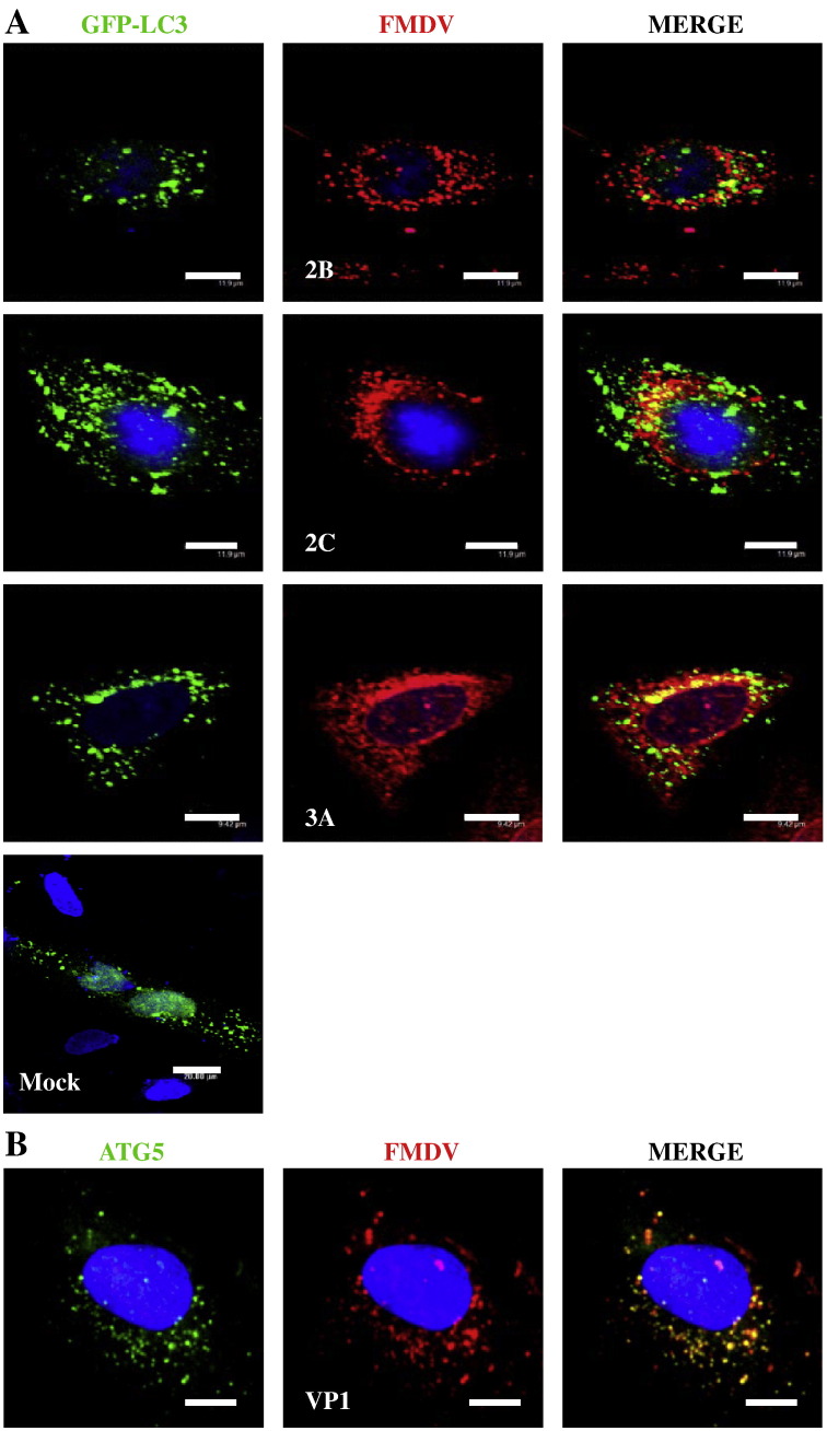
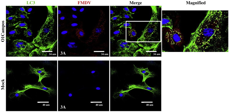
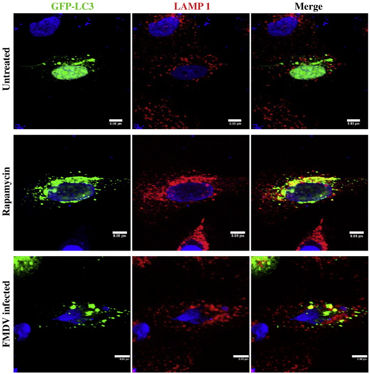
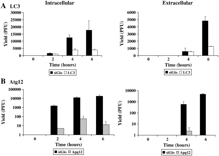
Foot-and-mouth disease virus (FMDV) is the type species of the Aphthovirus genus within the Picornaviridae family. Infection of cells with positive-strand RNA viruses results in a rearrangement of intracellular membranes into viral replication complexes. The origin of these membranes remains unknown; however induction of the cellular process of autophagy is beneficial for the replication of poliovirus, suggesting that it might be advantageous for other picornaviruses. By using confocal microscopy we showed in FMDV-infected cells co-localization of non-structural viral proteins 2B, 2C and 3A with LC3 (an autophagosome marker) and viral structural protein VP1 with Atg5 (autophagy-related protein), and LC3 with LAMP-1. Importantly, treatment of FMDV-infected cell with autophagy inducer rapamycin, increased viral yield, and inhibition of autophagosomal pathway by 3-methyladenine or small-interfering RNAs, decreased viral replication. Altogether, these studies strongly suggest that autophagy may play an important role during the replication of FMDV.


Figures

Fig. 1
Fig. 1
Analysis of redistribution of autophagy marker GFP–LC3 and FMDV proteins in MCF-10A cells. A. MCF-10A cells were transfected with a plasmid that expresses a GFP–LC3 (green) fusion protein as indicated. Forty-eight hours after transfection, cells were infected with FMDV O1Campos or mock infected and processed for immunofluorescence staining as described in Materials and methods. FMDV non-structural proteins were detected with specific MAbs and visualized with Alexa Fluor 594 (red). B. MCF-10A cells were infected with FMDV O1Campos and processed for immunofluorescence staining as describe in Materials and methods. FMDV viral capsid protein VP1, was detected with a MAb and visualized with Alexa Fluor 594 (red); Atg5 was detected with a rabbit antibody and visualized with Alexa Fluor 488 (green).
Fig. 2
Fig. 2
Analysis of redistribution of autophagy marker LC3 and non-structural FMDV protein 3A in bovine pharynx epithelial primary cultures. Primary cultures of bovine pharynx were infected with FMDV O1Campos or mock infected and processed for immunofluorescence staining as described in Materials and methods. FMDV non-structural protein 3A was detected with a MAb and visualized with Alexa Fluor 594 (red). LC3 was detected with a rabbit antibody and visualized with Alexa Fluor 488 (green).
Fig. 3
Fig. 3
Detection of GFP–LC3 and LAMP-1 in FMDV-infected cells and in the presence of an autophagy inducer, rapamycin. MCF-10A cells were transfected with a plasmid that expresses a GFP–LC3 (green) fusion protein as indicated. Forty-eight hours after transfection, cells were mock infected or infected with FMDV O1Campos, or treated with rapamycin. Cells were processed for immunofluorescence staining as described in Materials and methods. LAMP-1 was detected with a MAb and visualized with Alexa Fluor 594 (red).
Fig. 4
Fig. 4
Viral yields from FMDV-infected cells in the presence of the inducer of autophagy, rapamycin, or the inhibitor of autophagy, 3-MA. MCF-10A cells (A) or bovine pharynx epithelial primary cultures (B) were pre-treated with rapamycin (50 nM) or 3-MA (20 mM) for 2 h before infection with FMDV O1Campos at an MOI 1, as described in Materials and methods. Viral yields were determined by plaque assay in BHK-21 cells and represented as the ratio of plaque titers of treated and untreated infected cells at 5 hpi. Values are averages and standard deviations representative of two independent experiments.
Fig. 5
Fig. 5
Viral yields from FMDV infections in MCF-10A cells treated with siRNA to reduce the intracellular concentration of LC3 and Atg12. MCF-10A cells were transfected with four RNA duplexes targeted to LC3 (A), Atg12 (B) or with a control siRNA, siGlo for 48 h at 37 °C as described in Materials and methods. After transfection triplicate plates were infected with FMDV O1Campos at an MOI of 1 PFU/cell for the indicated times. Plaque titers were determined in BHK-21 cells and expressed as PFU/ml for the intra- and extracellular virus.
Fig. 6
Fig. 6
A — Electron micrographs of mock- and FMDV-infected MCF-10A cells. B — Immunogold detection of LAMP-1 and FMDV 2B non-structural protein individually. LAMP-1 and FMDV 2B proteins were visualized with specific MAb and detected with anti-mouse conjugated with ultra-small gold particles. Note that both proteins are found associated with the infection associated-membranes in the infected cells.

References

    1. Alexandersen S., Zhang Z., Donaldson E.I. Aspects of the persistence of foot-and-mouth disease virus in animals — the carrier problem. Microbes Infect. 2002;4:1099–1110. - PubMed
    1. Armer H., Moffat K., Wileman T., Belsham G.J., Jackson T., Duprex W.P., Ryan M., Monaghan P. Foot-and-mouth disease virus, but not Bovine enterovirus, targets the host cell cytoskeleton via the nonstructural protein 3Cpro. J. Virol. 2008;82(21):10556–10566. - PMC - PubMed
    1. Arzt J., Pacheco J.M., Rodriguez L.L. The early pathogenesis of foot-and-mouth disease in cattle after aerosol inoculation: identification of the nasopharynx as the primary site of infection. Vet. Pathol. 2010 - PubMed
    1. Barlow Y., Pye R.J. Keratinocyte culture. In: Pollard J.W., Waljer J.M., editors. Methods in Molecular Biology. vol. 5. Humana Press; Clifton, NJ: 1989. pp. 83–98. (Animal Cell Culture). - PubMed
    1. Bienz K., Egger D., Wolff D.A. Virus replication, cytopathology, and lysosomal enzyme response of mitotic and interphase hep-2 cells infected with poliovirus. J. Virol. 1973;11(4):565–574. - PMC - PubMed

Publication types