Review
. 2011 Feb;21(1):32-41.
doi: 10.1016/j.sbi.2010.11.001. Epub 2010 Nov 26.

Protein folding in the cell: challenges and progress

Affiliations
Review

Protein folding in the cell: challenges and progress

Anne Gershenson et al. Curr Opin Struct Biol. 2011 Feb.

Abstract

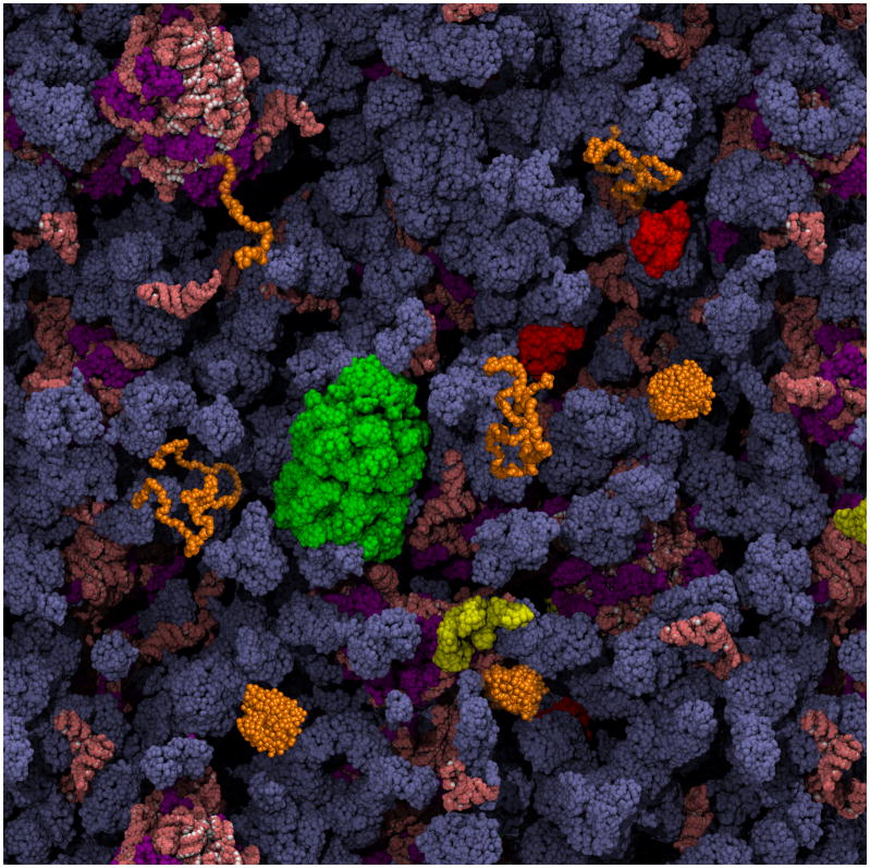
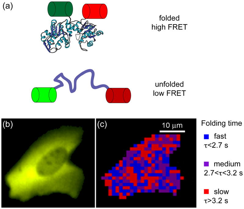
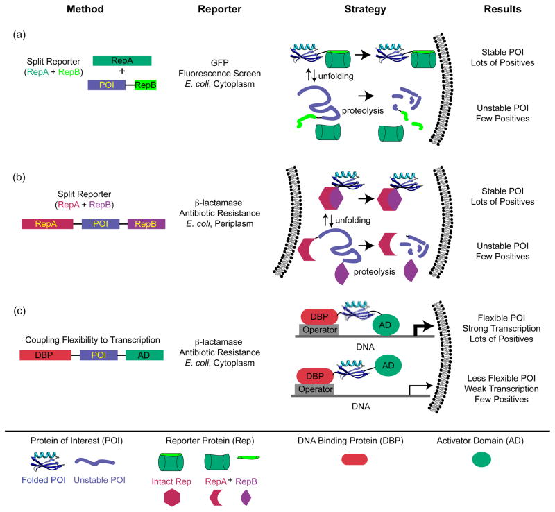
It is hard to imagine a more extreme contrast than that between the dilute solutions used for in vitro studies of protein folding and the crowded, compartmentalized, sticky, spatially inhomogeneous interior of a cell. This review highlights recent research exploring protein folding in the cell with a focus on issues that are generally not relevant to in vitro studies of protein folding, such as macromolecular crowding, hindered diffusion, cotranslational folding, molecular chaperones, and evolutionary pressures. The technical obstacles that must be overcome to characterize protein folding in the cell are driving methodological advances, and we draw attention to several examples, such as fluorescence imaging of folding in cells and genetic screens for in-cell stability.


Figures

Figure 1
Figure 1. Schematic depiction of a protein folding reaction in the cytoplasm of an E. coli cell, showing vividly how different the environment is from dilute in vitro refolding experiments
The cytoplasmic components are present at their known concentrations. As discussed in this review, features of particular importance to the folding of a protein of interest (in orange) are: the striking extent of volume exclusion due to macromolecular crowding, the presence of molecular chaperones that interact with nascent and incompletely folded proteins (GroEL in green, DnaK in red, and trigger factor in yellow), and the possibility of co-translational folding upon emergence of the polypeptide chain from the ribosome (ribosomal proteins are purple; all RNA is salmon). The cytoplasm image is courtesy of A. Elcock.
Figure 2
Figure 2. Monitoring protein folding kinetics in a living cell using fast relaxation imaging (FReI) [68]
(a) The folding sensor was created by sandwiching a temperature-sensitive POI (here, PGK) between two fluorescent proteins, GFP and mCherry, allowing folding to be monitored by FRET. (b) Image of the fluorescence arising from expression of the fusion protein in a human cell. (c) Map of the refolding dynamics of the fusion protein in the cell shown in (b). The time dependence of the FRET signal following a temperature jump is spatially mapped. Figures 2b and c are courtesy of M. Gruebele.
Figure 3
Figure 3. Selection and screening strategies designed to assess protein stability in vivo
(a) Construct designed such that GFP fluorescence reports on protein solubility, which is correlated to stability [74]. The gene for the POI is C-terminally fused to a β strand from GFP, and a GFP construct missing this strand is co-expressed with the fusion. GFP fluorescence reports on the successful docking of the missing strand onto the truncated GFP. In turn, unstable (or insoluble) POIs with their attached GFP strands are degraded (or inaccessible to recombine with the truncated GFP). (b) Screen designed such that successful periplasmic expression of β-lactamase requires a stably folded POI and hence recombination of two β-lactamase fragments that flank the POI [75]. Here, the level of functional periplasmic β-lactamase, which relies on stability of the POI, is read out as resistance to increasing concentrations of β–lactam antibiotic [75]. As in (a), this correlation is presumably based on the enhanced proteolytic susceptibility of the fusion protein when the POI is unstable. (c) Construct designed such that flexibility of the POI controls transcription of the β-lactamase gene [78,79]. When the POI is flanked by the N-terminal DNA-binding domain of bacteriophage λ, which binds to the λ operator, and the RNA polymerase α subunit, which activates transcription of the β-lactamase gene, flexible POIs lead to greater transcriptional activation and higher antibiotic resistance.

References

    1. Zhou HX, Rivas G, Minton AP. Macromolecular crowding and confinement: biochemical, biophysical, and potential physiological consequences. Annu Rev Biophys. 2008;37:375–397. - PMC - PubMed
    1. Elcock AH. Models of macromolecular crowding effects and the need for quantitative comparisons with experiment. Curr Opin Struct Biol. 2010;20:196–206. - PMC - PubMed
    1. Tsao D, Dokholyan NV. Macromolecular crowding induces polypeptide compaction and decreases folding cooperativity. Phys Chem Chem Phys. 2010;12:3491–3500. - PMC - PubMed
    1. Hong J, Gierasch LM. Macromolecular crowding remodels the energy landscape of a protein by favoring a more compact unfolded state. J Am Chem Soc. 2010;132:10445–10452. - PMC - PubMed
    1. Wang Y, He H, Li S. Effect of Ficoll 70 on thermal stability and structure of creatine kinase. Biochemistry. 2010;75:648–654. - PubMed

Publication types

LinkOut - more resources