. 2011 Feb;300(2):L255-65.
doi: 10.1152/ajplung.00304.2010. Epub 2010 Nov 26.

Cigarette smoke-induced lung emphysema in mice is associated with prolyl endopeptidase, an enzyme involved in collagen breakdown

Affiliations

Cigarette smoke-induced lung emphysema in mice is associated with prolyl endopeptidase, an enzyme involved in collagen breakdown

Saskia Braber et al. Am J Physiol Lung Cell Mol Physiol. 2011 Feb.

Abstract

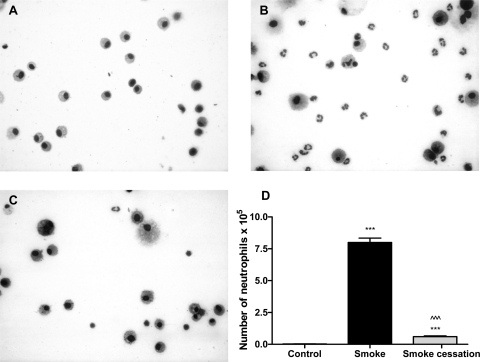
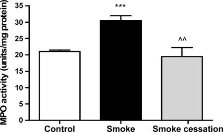
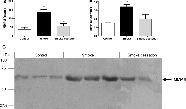
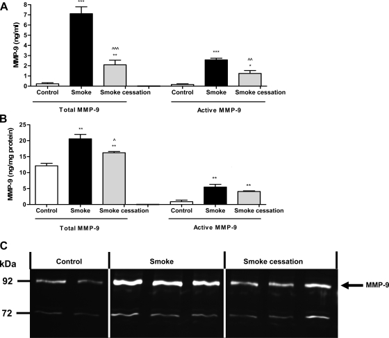
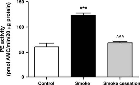
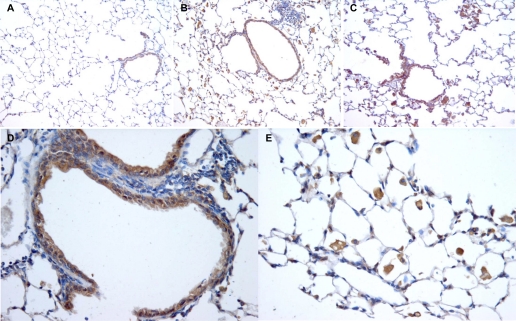
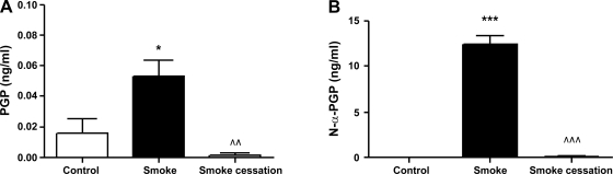
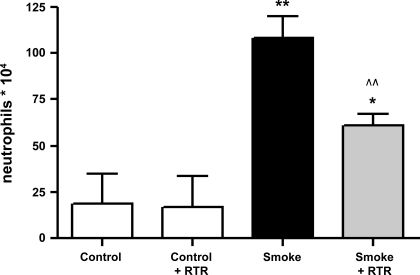
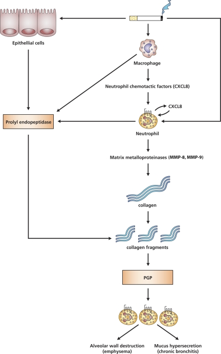
There is increasing evidence that the neutrophil chemoattractant proline-glycine-proline (PGP), derived from the breakdown of the extracellular matrix, plays an important role in neutrophil recruitment to the lung. PGP formation is a multistep process involving neutrophils, metalloproteinases (MMPs), and prolyl endopeptidase (PE). This cascade of events is now investigated in the development of lung emphysema. A/J mice were whole body exposed to cigarette smoke for 20 wk. After 20 wk or 8 wk after smoking cessation, animals were killed, and bronchoalveolar lavage fluid and lung tissue were collected to analyze the neutrophilic airway inflammation, the MMP-8 and MMP-9 levels, the PE activity, and the PGP levels. Lung tissue degradation was assessed by measuring the mean linear intercept. Additionally, we investigated the effect of the peptide L-arginine-threonine-arginine (RTR), which binds to PGP sequences, on the smoke-induced neutrophil influx in the lung after 5 days of smoke exposure. Neutrophilic airway inflammation was induced by cigarette smoke exposure. MMP-8 and MMP-9 levels, PE activity, and PGP levels were elevated in the lungs of cigarette smoke-exposed mice. PE was highly expressed in epithelial and inflammatory cells (macrophages and neutrophils) in lung tissue of cigarette smoke-exposed mice. After smoking cessation, the neutrophil influx, the MMP-8 and MMP-9 levels, the PE activity, and the PGP levels were decreased or reduced to normal levels. Moreover, RTR inhibited the smoke-induced neutrophil influx in the lung after 5 days' smoke exposure. In the present murine model of cigarette smoke-induced lung emphysema, it is demonstrated for the first time that all relevant components (neutrophils, MMP-8, MMP-9, PE) involved in PGP formation from collagen are upregulated in the airways. Together with MMPs, PE may play an important role in the formation of PGP and thus in the pathophysiology of lung emphysema.


Figures

Fig. 1.
Fig. 1.
Neutrophil influx in the bronchoalveolar lavage (BALF) is related to smoke exposure. Representative photomicrographs of DiffQuick-stained cytospins on glass slides of BALF from air-exposed mice (A), smoke-exposed mice (B), and smoke-exposed mice 8 wk after smoke cessation (C). Magnification, ×1000. D: absolute neutrophil numbers in the BALF of mice exposed to air, mice exposed to cigarette smoke for 20 wk (black bar), and mice exposed to cigarette smoke for 20 wk + a smoking cessation period of 8 wk (gray bar). N = 4–5 animals/group. Values are expressed as means ± SE. ***P ≤ 0.001; significantly different from the control group. ^^^P ≤ 0.001; significantly different from the smoke group.
Fig. 2.
Fig. 2.
Elevated MPO activity in lung homogenates after chronic smoke exposure. MPO activity in lung homogenates of mice exposed to air (white bar), mice exposed to cigarette smoke for 20 wk (black bar), and mice exposed to cigarette smoke for 20 wk + a smoking cessation period of 8 wk (gray bar). N = 4–6 animals/group. Values are expressed as means ± SE. ***P ≤ 0.001; significantly different from the control group. ^^P ≤ 0.01; significantly different from the smoke group.
Fig. 3.
Fig. 3.
Smoke-related MMP-8 increase in BALF. The total MMP-8 levels were determined in the BALF (A) after 20-wk air exposure (white bar), after 20-wk smoke exposure (black bar), and after 20-wk smoke exposure + a smoking cessation period of 8 wk (gray bar) via ELISA. N = 4–5 animals/group. Values are expressed as means ± SE. **P ≤ 0.01; significantly different from the control group. ^^P ≤ 0.01; significantly different from the smoke group. BALF samples from 3 control mice, 3 smoke-exposed mice, and 2 smoke-exposed mice after a smoking cessation period were randomly chosen and analyzed via Western blotting (C). The molecular weight bands at ∼65 kDa likely represent active MMP-8, and the optical density of these molecular weight bands was measured (B). The position of the bands for MMP-8 is indicated by an arrow.
Fig. 4.
Fig. 4.
Smoke-related MMP-9 increase in BALF and lung homogenates. The total and active MMP-9 levels were determined in the BALF (A) and lung homogenates (B) after 20-wk air exposure (white bars), after 20-wk smoke exposure (black bars), and after 20-wk smoke exposure + a smoking cessation period of 8 wk (gray bars). N = 4–5 animals/group. Values are expressed as means ± SE. *P ≤ 0.05, **P ≤ 0.01, ***P ≤ 0.001; significantly different from the control group. ^P ≤ 0.05, ^^P ≤ 0.01, ^^^P ≤ 0.001; significantly different from the smoke group. Lung homogenates from 2 control mice, 3 smoke-exposed mice, and 3 smoke-exposed mice after a smoking cessation period were randomly chosen and analyzed by gelatin zymography (C). Gelatinolytic activity of MMP-9 was increased in the lung homogenates after 20 wk of smoke exposure compared with the air-exposed mice, whereas after smoking cessation, the activity of MMP-9 was decreased. The position of the bands for MMP-9 is indicated by an arrow.
Fig. 5.
Fig. 5.
Prolyl endopeptidase (PE) activity was elevated in lung homogenates after chronic smoke exposure. PE activity in lung homogenates of mice exposed to air (white bar), mice exposed to cigarette smoke for 20 wk (black bar), and mice exposed to cigarette smoke for 20 wk + a smoking cessation period of 8 wk (gray bar). N = 4–5 animals/group. Values are expressed as means ± SE. ***P ≤ 0.001; significantly different from the control group. ^^^P ≤ 0.001; significantly different from the smoke group.
Fig. 6.
Fig. 6.
Localization of PE in the lung. Representative photomicrographs of an immunohistological staining for PE (brown color, DAB staining) in lung tissue of air-exposed mice (A), smoke-exposed mice (B), and smoke-exposed mice after a smoking cessation period of 8 wk (C). Representative photomicrographs of lung tissue of a smoke-exposed mouse with a pronounced expression of PE in the epithelial cells (D) and the inflammatory cells (E). Magnification, ×200 (A–C), ×400 (D and E). N = 3 animals/group.
Fig. 7.
Fig. 7.
Proline-glycine-proline (PGP) and N-α-PGP were detectable in BALF after chronic smoke exposure. The PGP (A) and N-α-PGP (B) levels were determined in the BALF after 20-wk air exposure (white bars), after 20-wk smoke exposure (black bars), and after 20-wk smoke exposure + a smoking cessation period of 8 wk (gray bars). N = 4–5 animals/group. Values are expressed as means ± SE. *P ≤ 0.05, ***P ≤ 0.001; significantly different from the control group. ^^P ≤ 0.01, ^^^P ≤ 0.001; significantly different from the smoke group.
Fig. 8.
Fig. 8.
Cigarette smoke-induced neutrophil influx in the BALF was significantly decreased after l-arginine-threonine-arginine (RTR) administration. Absolute neutrophil numbers in the BALF of mice exposed to air, RTR-treated mice exposed to air, mice exposed to cigarette smoke, and RTR-treated mice exposed to cigarette smoke for 5 days. The mice received vehicle (PBS) or RTR by oropharyngeal aspiration (50 μg/50 μl PBS) twice daily. N = 4–6 animals/group. Values are expressed as means ± SE. *P ≤ 0.05, **P ≤ 0.01; significantly different from the control group. ^^P ≤ 0.01; significantly different from the smoke group.
Fig. 9.
Fig. 9.
PGP generation is a multistep process. In lung emphysema, the cascade of events leading to PGP formation will start with cigarette smoke exposure. Cigarette smoke can stimulate alveolar macrophages to release several chemoattractants, such as CXCL8. Subsequently, CXCL8 facilitates the migration of neutrophils to the site of inflammation. The activated neutrophils are also capable themselves to produce CXCL8, and the cigarette smoke exposure will also affect these neutrophils. PGP formation is a multistep process initially involving release of proteases from the MMP family, like MMP-8 and MMP-9. MMP-8 and MMP-9 are released by activated neutrophils and can proteolytically cleave collagen to smaller fragments resulting in an optimal substrate for PE activity. These collagen fragments are then further cleaved to PGP by PE, a member of the serine protease family. Various cell types, like neutrophils, macrophages, and epithelial cells, express PE. The generated PGP is chemotactic for neutrophils and results in an environment of chronic inflammation with proteolytic damage and PGP formation. Finally, this will lead to alveolar wall destruction (emphysema) and mucus hypersecretion (chronic bronchitis).

References

    1. Abboud RT, Vimalanathan S. Pathogenesis of COPD. Part I. The role of protease-antiprotease imbalance in emphysema. Int J Tuberc Lung Dis 12: 361–367, 2008 - PubMed
    1. Atkinson JJ, Senior RM. Matrix metalloproteinase-9 in lung remodeling. Am J Respir Cell Mol Biol 28: 12–24, 2003 - PubMed
    1. Barnes PJ. Chronic obstructive pulmonary disease. N Engl J Med 343: 269–280, 2000 - PubMed
    1. Barrett AJ, Rawlings ND. Oligopeptidases, and the emergence of the prolyl oligopeptidase family. Biol Chem Hoppe Seyler 373: 353–360, 1992 - PubMed
    1. Belvisi MG, Bottomley KM. The role of matrix metalloproteinases (MMPs) in the pathophysiology of chronic obstructive pulmonary disease (COPD): a therapeutic role for inhibitors of MMPs? Inflamm Res 52: 95–100, 2003 - PubMed

Publication types

MeSH terms

LinkOut - more resources