. 2011 Jan 5;30(1):221-31.
doi: 10.1038/emboj.2010.297. Epub 2010 Nov 26.

Metalloprotease type III effectors that specifically cleave JNK and NF-κB

Affiliations

Metalloprotease type III effectors that specifically cleave JNK and NF-κB

Kobi Baruch et al. EMBO J. .

Abstract

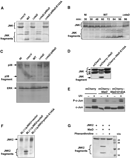
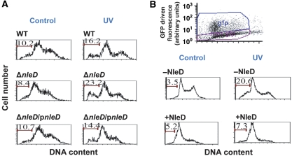
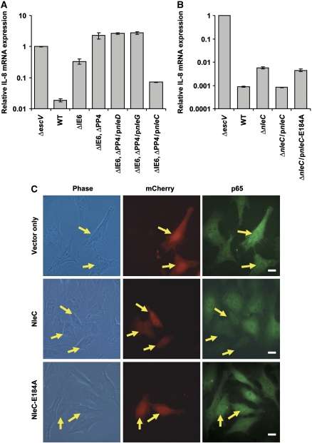
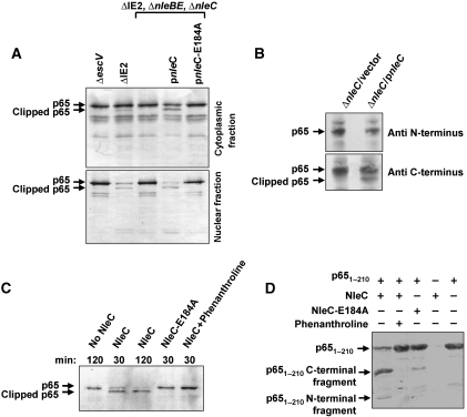
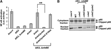
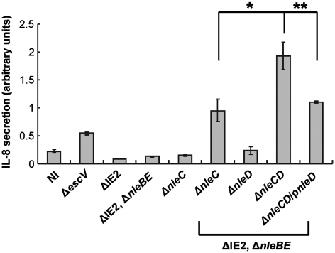
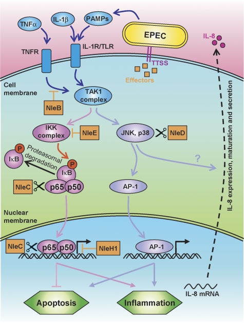
Two major arms of the inflammatory response are the NF-κB and c-Jun N-terminal kinase (JNK) pathways. Here, we show that enteropathogenic Escherichia coli (EPEC) employs the type III secretion system to target these two signalling arms by injecting host cells with two effector proteins, NleC and NleD. We provide evidence that NleC and NleD are Zn-dependent endopeptidases that specifically clip and inactivate RelA (p65) and JNK, respectively, thus blocking NF-κB and AP-1 activation. We show that NleC and NleD co-operate and complement other EPEC effectors in accomplishing maximal inhibition of IL-8 secretion. This is a remarkable example of a pathogen using multiple effectors to manipulate systematically the host inflammatory response signalling network.


Conflict of interest statement

The authors declare that they have no conflict of interest.

Figures

Figure 1
Figure 1
Involvement of NleD in JNK cleavage. (A) NleD is required for JNK degradation. HeLa cells were infected with one of the following EPEC: wild-type (WT), nleD deletion mutant (ΔnleD), nleD deletion mutant complemented with a plasmid expressing wild-type nleD (pKB4345, indicated as pNleD) or nleD deletion mutant complemented with a plasmid expressing mutated nleD (pLG4457, indicated as pNleD-E143A). After 3 h, proteins were extracted and subjected to western blot analysis using anti-JNK antibody. JNK and its degradation fragments are indicated. Cells infected with a TTSS-deficient mutant (ΔescV) were used as negative control. (B) The kinetics of JNK cleavage upon EPEC infection. HeLa cells were infected with EPEC primed to express the TTSS for the indicated periods before proteins were extracted and subjected to western blot analysis using anti-JNK antibody. JNK and its degradation fragments are indicated. Non-infected cells (NI) and cells infected with nleD deletion mutant (ΔnleD) served as controls. (C) NleD induces cleavage of p38, but not that of ERK. HeLa cells were infected as indicated in (A). After 3 h, proteins were extracted and subjected to western blot analysis using anti-p38 or anti-ERK antibodies. ERK, p38 and its degradation fragments are indicated. (D) Ectopically expressed NleD correlates with JNK degradation. HEK293 cells were transfected with one of the following plasmids expressing mCherry–NleD, mCherry–NleD-E143A, or mCherry (pLG4419, pLG4477, and pSC4141, respectively). After 24 h, proteins were extracted and subjected to western blot analysis using anti-JNK antibody. JNK and its degradation fragments are indicated. (E) Ectopically expressed NleD is associated with inhibition JNK activity. HEK293 cells transfected with plasmids expressing mCherry, mCherry–NleD, or mCherry–NleD-E143A, were irradiated with 30 J/m2 of UV and harvested 3 h later. The levels of phospho-c-Jun and total c-Jun were determined by western analysis using anti-c-Jun and anti-phospho-c-Jun antibodies. (F) NleD induces cleavage of JNK in E. coli cytoplasm. E. coli BL21 was co-transformed with plasmid expressing JNK2 and either vector only (pCX341) or plasmid expressing nleD (pEM3654). Co-expression of JNK2 and NleD was induced for 2 h by IPTG, before proteins were extracted and subjected to western blot analysis using anti-JNK antibody. JNK and its degradation fragments are indicated. (G) NleD clips JNK in vitro. Purified JNK2 and NleD were incubated in a reaction mixture at a molar ratio of 40:1, in the presence or absence of the Zn protease inhibitor phenanthroline. The reaction was stopped by addition of SDS loading buffer and proteins separated by SDS–PAGE. Finally, proteins were visualized by Coomassie blue staining. JNK2 and its degradation fragments are indicated. Of note, NleD does not appear in this gel as its concentration is below detection levels.
Figure 2
Figure 2
NleD clips JNK within the activation loop. (A) Schematic diagram of JNK2. The location of the HA and anti-JNK antibody epitopes, activation loop (A loop), T and Y phosphorylation sites and NleD-cleavage site are indicated. (B, C) NleD cleaves JNK2 in vivo. HeLa cells were transfected with plasmids expressing HA-tagged JNK2. At 25 h post-transfection, the cells were infected with wild-type (WT) or nleD deletion mutant (ΔnleD) EPEC. After 2.5 h, proteins were extracted from the infected HeLa cells, immunoprecipitated using anti-HA antibody and subjected to western blot analysis using anti-HA (B) or anti-JNK (C) antibodies. JNK2 and its degradation fragments are indicated. Negative control using non-transfected cells confirmed the antibody specificity (data not shown). (D, E) NleD cleaves JNK2 in vitro. Purified, N-terminally tagged, 6His-JNK2 was incubated with purified NleD or NleD-E143A. After 60 min, the reaction was stopped using SDS loading buffer. To estimate the size of the N-terminal fragment of the clipped JNK2, the reaction mixture was subjected to western blot analysis using anti-6His antibody (D). Intact JNK2 and its N-terminal fragments are indicated. To determine the size of the two JNK2 fragments, the reaction mixture was analysed also by SDS–PAGE followed by Commassie blue staining (E). JNK2 and its degradation fragments are indicated. The framed C-terminal band was subjected to N-terminal sequencing analysis.
Figure 3
Figure 3
NleD inhibits JNK-dependent apoptosis. (A) HeLa cells were infected with wild-type EPEC, nleD mutant and complemented mutant. After infection the cells were washed, UV irradiated (30 J/m2), and harvested 4 h later. The DNA was stained with propidium iodide and cells were analysed by flow cytometry. Cells containing sub-G1 DNA are indicated by bars and their percentage in the population is indicated above the bars. Irradiated cells are indicated by ‘UV' and non-irradiated control cells by ‘Control'. (B) RKO cells were transfected with plasmid expressing NleD–GFP fusion protein and UV irradiated 24 h later (30 J/m2), or not (marked as Control). After additional 12 h, cells were harvested and analysed by flow cytometry. The non-transfected population (marked as −NleD), and the GFP expressing, transfected cells (+NleD), were gated, as exhibited in the upper panel, and the DNA content in each gated population was determined. Cells containing sub-G1 DNA are indicated by bars and their percentage in the population is indicated above the bars.
Figure 4
Figure 4
NleC expression correlates with inhibition of the NF-κB pathway. (A) Deletion analysis to identify the EPEC gene that represses IL-8 induction. HeLa cells were infected with one of the following EPEC: EPEC with a deleted IE6 region (ΔIE6), EPEC with deleted IE6 and PP4 regions (ΔIE6 and ΔPP4), or the latter complemented with plasmids expressing either wild-type NleD, NleG, or NleC (pnleD, pnleG, and pnleC, respectively). Cells infected with TTSS mutant (ΔescV) and wild-type EPEC (WT) served as negative and positive controls, respectively. HeLa cells were infected with the relevant EPEC for 2 h to allow injection of effectors before stimulation with TNFα for 3 h. Then RNA was extracted from the HeLa cells and real-time PCR performed to quantify IL-8 mRNA levels. Experiments were performed in duplicates and a typical experiment out of three is shown. Error bars indicate s.d. (B) NleC is required for inhibition of TNFα-induced IL-8 expression. HeLa cells were infected with nleC mutant, or this mutant complemented with plasmids expressing wild-type NleC (pnleC) or NleC-E184A mutant (pnleC-E184A). TTSS mutant (ΔescV) or wild-type EPEC (WT) served as negative and positive controls, respectively. HeLa cells were infected with these strains as described in (A), RNA was extracted from the HeLa cells and real-time PCR performed to quantify IL-8 mRNA levels. The indicated values are relative to IL-8 RNA levels in cells infected with ΔescV mutant. Experiments were performed in duplicates and a typical experiment out of four is shown. Error bars indicate the s.d. (C) NleC reduces p65 levels in vivo. HeLa cells transfected with plasmid expressing mCherry, mCherry–NleC or mCherry–NleC-E184A (red) were treated with TNFα for 30 min, after which they were fixed and visualized using anti-p65 antibody (green). Yellow arrows indicate cells expressing mCherry proteins. Bar represents 20 μm.
Figure 5
Figure 5
NleC induces p65 cleavage, which is associated with reduced nuclear p65 levels. (A) NleC induces p65 cleavage in vivo. HeLa cells were infected for 3 h with the ΔIE2 ΔnleBE, ΔnleC mutant EPEC that was complemented, or not, with plasmids expressing NleC or mutated NleC (NleC-E184A), as indicated. Proteins were extracted from the infected HeLa cells, separated into cytosolic and nuclear fractions and subjected to western blot analysis using anti-p65 antibody. An EPEC mutant deficient in TTSS activity (ΔescV) and EPEC deleted of the IE2 region (ΔIE2) served as negative and positive controls, respectively; the latter was used here as wild type. Intact and clipped p65 are indicated. (B) NleC induces clipping the N-terminal end of p65. HeLa cells were infected for 3 h with the ΔnleC mutant EPEC that was complemented with plasmids expressing NleC or vector only, as indicated. Proteins were extracted from the infected HeLa cells and subjected to western blot analysis using anti-N-terminus of p65 or anti-C-terminus of p65 antibodies. Intact and clipped p65 are indicated. (C) NleC induced p65 cleavage in vitro. The cytosolic fraction of HeLa extracts was combined with NleC, NleC-E184A, or buffer alone in the presence or absence of phenanthroline, a Zn metalloprotease inhibitor, as indicated. The incubation time is indicated above each lane. Reaction products were visualized by western blot analysis using anti-p65 antibody. Intact and clipped p65 are indicated. (D) NleC directly cuts p651–210. Purified p651–210 and NleC were incubated in a reaction mixture at a molar ratio of 40:1, in the presence or absence of phenanthroline. The reaction was stopped by addition of SDS loading buffer and proteins separated by SDS–PAGE and visualized by Coomassie blue staining. p651–210 and its degradation fragments are indicated. NleC does not appear in this gel.
Figure 6
Figure 6
Interplay between NleC and NleBE, in mediating NF-κB repression. (A) NleC and NleBE are required for complete repression of NF-κB. HeLa cells were transfected with a mix of plasmid expressing luc via an NF-κB promoter and a plasmid expressing renilla-luc expressed from constitutive promoter. The cells were next infected with different EPEC strains, and after 3 h, infection was stopped by replacing the media with DMEM supplemented with gentamicin. At 7 h post-infection, the levels of luc and renilla-luc expression were determined. Shown is a reprehensive experiment out of two that gave similar results. The assay was performed in triplicates. Error bars indicate the s.d. (**P-value<0.05). (B) NleBE expression is associated with attenuation the NleC-dependent p65 cleavage. HeLa cells were infected with various EPEC strains as indicated. After 3 h, proteins were extracted, separated to cytoplasmic and nuclear fractions and subjected to western analysis using anti-p65 C-terminal antibody. The intact and clipped p65 are indicated.
Figure 7
Figure 7
NleE, NleB, NleC, and NleD are required for maximal EPEC-induced repression of IL-8 secretion. HeLa cells were infected with different EPEC strains as indicated. After 3 h, cells were washed and the media was replaced by DMEM supplemented with gentamicin. At 19 h post-infection, the media was harvested from the respective wells, cleared and the amount of secreted IL-8 determined by ELISA. The relative amounts of secreted IL-8 are shown. Experiments were performed in triplicates and a typical experiment out of two is shown. Standard deviation (bars) (*P-value<0.1, **P-value<0.05).
Figure 8
Figure 8
A model of the anti-inflammatory activity of NleBCDE and NleH1. Schematic diagram of the signalling cascades activated by PAMPs, IL-1, and TNF is shown. The different EPEC effectors (orange boxes) interact with this signalling network at multiple points. NleB inhibits the TNFR signalling upstream to the TAK1 complex, NleE prevents IKK activation, NleC cleaves and inactivates cytoplasmic and nuclear p65, and NleH1 inhibits the interaction of NF-κB with some promoters. Finally, NleD cuts and inactivates JNK and p38. These effectors function in concert to repress IL-8 secretion and to modulate the inflammation and apoptosis processes.

References

    1. Auerbuch V, Golenbock DT, Isberg RR (2009) Innate immune recognition of Yersinia pseudotuberculosis type III secretion. PLoS Pathog 5: e1000686. - PMC - PubMed
    1. Barkett M, Gilmore TD (1999) Control of apoptosis by Rel/NF-kappaB transcription factors. Oncogene 18: 6910–6924 - PubMed
    1. Chen FE, Huang DB, Chen YQ, Ghosh G (1998a) Crystal structure of p50/p65 heterodimer of transcription factor NF-kappaB bound to DNA. Nature 391: 410–413 - PubMed
    1. Chen YQ, Ghosh S, Ghosh G (1998b) A novel DNA recognition mode by the NF-kappa B p65 homodimer. Nat Struct Biol 5: 67–73 - PubMed
    1. Chen ZJ (2005) Ubiquitin signalling in the NF-kappaB pathway. Nat Cell Biol 7: 758–765 - PMC - PubMed

Publication types

MeSH terms