. 2011 Jan 6;469(7328):47-52.
doi: 10.1038/nature09635. Epub 2010 Nov 28.

Reversing EphB2 depletion rescues cognitive functions in Alzheimer model

Affiliations

Reversing EphB2 depletion rescues cognitive functions in Alzheimer model

Moustapha Cissé et al. Nature. .

Abstract

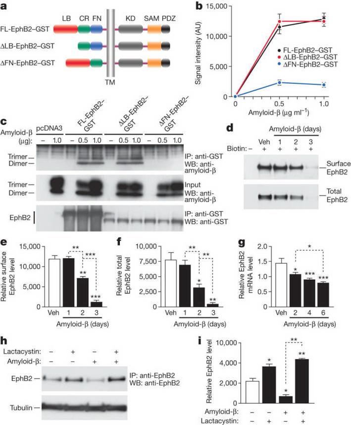
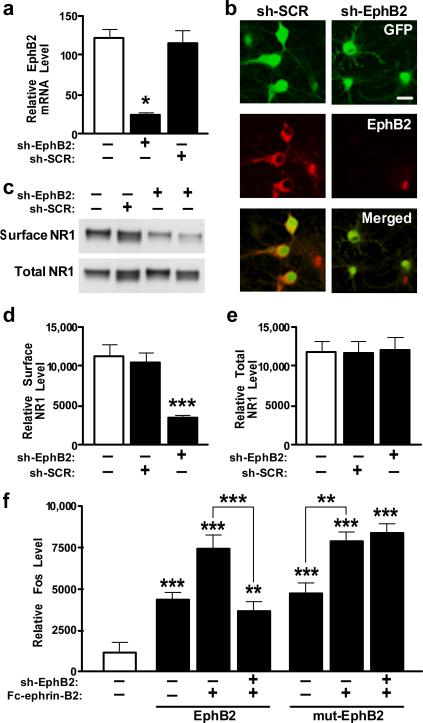
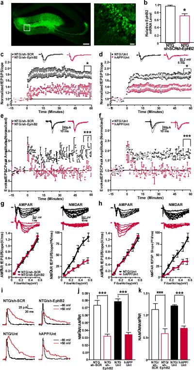
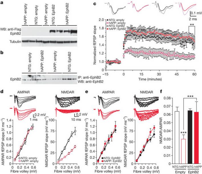
Amyloid-β oligomers may cause cognitive deficits in Alzheimer's disease by impairing neuronal NMDA-type glutamate receptors, whose function is regulated by the receptor tyrosine kinase EphB2. Here we show that amyloid-β oligomers bind to the fibronectin repeats domain of EphB2 and trigger EphB2 degradation in the proteasome. To determine the pathogenic importance of EphB2 depletions in Alzheimer's disease and related models, we used lentiviral constructs to reduce or increase neuronal expression of EphB2 in memory centres of the mouse brain. In nontransgenic mice, knockdown of EphB2 mediated by short hairpin RNA reduced NMDA receptor currents and impaired long-term potentiation in the dentate gyrus, which are important for memory formation. Increasing EphB2 expression in the dentate gyrus of human amyloid precursor protein transgenic mice reversed deficits in NMDA receptor-dependent long-term potentiation and memory impairments. Thus, depletion of EphB2 is critical in amyloid-β-induced neuronal dysfunction. Increasing EphB2 levels or function could be beneficial in Alzheimer's disease.


Figures

Figure 1
Figure 1. Amyloid-β oligomers bind to the fibronectin repeats domain of EphB2 and cause degradation of EphB2 in the proteasome
a, Domain structure of full-length (FL) EphB2 and deletion constructs. Ligand-binding (LB) domain, cysteine-rich (CR) region, fibronectin type III repeats (FN) domain, transmembrane (TM) region, tyrosine kinase (KD) domain, sterile alpha motif (SAM) domain, and PSD95, DLG and ZO1 (PDZ) domain. b, Binding of amyloid-β dimers and trimers to different EphB2 constructs. See Supplementary Table 2 for experimental details pertaining to data shown in figures. AU, arbitrary units. c, Representative western blot (WB). IP, immunoprecipitation. d–f, Amyloid-β-induced depletion of EphB2. Primary rat neurons were treated with amyloid-β or vehicle (Veh), and surface and total levels of EphB2 were determined by western blots. Representative western blots are shown in d. e, f, Quantification of surface (e) and total (f) levels of EphB2. g, EphB2 mRNA levels in primary neurons treated with amyloid-β or vehicle. h, i, Lactacystin blocks amyloid-β-induced depletion of EphB2 in primary neurons. Representative western blot (h) and quantification of signals (i). For all experiments, n = 3–6 wells per condition from three independent experiments. *P < 0.05, **P < 0.001, ***P < 0.0001 versus empty bars or as indicated by brackets (Tukey test). Values are means ± s.e.m.
Figure 2
Figure 2. Knockdown of EphB2 reduces surface NR1 levels and Fc-ephrin-B2-dependent Fos expression
a, b, EphB2 expression is reduced in primary neurons infected with Lenti-sh-EphB2–GFP as determined by RT–qPCR (a) or EphB2 immunostaining (b). Scale bar, 20 mm. c–e, Reduction of EphB2 levels by Lenti-sh-EphB2–GFP and effect on surface NR1 levels. f, shRNA against wild-type but not mutated EphB2 reduces Fc-ephrin-B2-dependent Fos expression. Primary rat neurons were co-infected or not with Lenti-sh-EphB2–GFP (sh-EphB2) in combination with either Lenti-EphB2 encoding wild-type EphB2 or Lenti-mut-EphB2 (mut-EphB2) encoding a mutated EphB2 mRNA that is not recognized by sh-EphB2. Four days later, cells were stimulated with clustered multimeric recombinant Fc-ephrin-B2 ligand to activate EphB2. n = 3–6 wells per condition from three independent experiments. *P < 0.05, **P < 0.001, ***P < 0.0001 versus empty bar or as indicated by brackets (Tukey's test). Values are means ± s.e.m.
Figure 3
Figure 3. Knockdown of EphB2 reduces LTP in dentate gyrus granule cells of nontransgenic mice
a, Anti-GFP immunostaining of dentate gyrus showing infected neurons in Lenti-sh-EphB2-GFP injected mice. Right panel shows higher magnification image of boxed region on left. Scale bars: 100 mm (left), 25 mm (right). b, EphB2 mRNA levels in the entire dentate gyrus (reflecting levels in infected and uninfected cells) (n = 5–7 mice per condition). *P < 0.001 versus sh-SCR (t test). NTG, nontransgenic mice. c–f, LTP at the medial perforant path to granule cell synapse measured by field recordings (c, d) or by whole-cell patch clamp from individual GFP-positive cells (e, f) in the dentate gyrus. LTP was impaired in nontransgenic mice treated with Lenti-sh-EphB2–GFP (sh-EphB2) compared to nontransgenic mice treated with Lenti-sh-SCR–GFP (sh-SCR) (c, e). Similar LTP impairments were observed in untreated (Unt) hAPP mice (d, f) (NTG: sh-EphB2 versus hAPP: Unt). *P < 0.05, ***P < 0.001 (repeated-measures ANOVA and Bonferroni post-hoc test on the last 10 min of data). n = 8–9 slices from 3–4 mice per treatment (c) or genotype (d). g, h, Comparison of AMPA-receptor (AMPAR)-mediated (left) and NMDA-receptor (NMDAR)-mediated (right) input-output (I/O) relationships in the medial perforant path to granule cell synapses of nontransgenic mice treated with sh-EphB2 versus sh-SCR (g) and of untreated nontransgenic (NTG: Unt) versus hAPP (hAPP: Unt) mice (h). i, Example traces of evoked glutamate receptor currents from individual granule cells voltage clamped at −80 or 50 mV to measure AMPA-receptor- and NMDA-receptor-mediated currents, respectively. j, k, Summary plot of the ratios of NMDA-receptor I/O relationships to AMPA-receptor I/O relationships measured by field recordings (i) or by individual granule cells (k). ***P < 0.001 (two-way ANOVA and Bonferroni post-hoc test). n = 8–9 slices from 3–4 mice per group. Values are means ± s.e.m.
Figure 4
Figure 4. Increasing EphB2 expression rescues synaptic plasticity in hAPP mice
a, b, Levels of EphB2–Flag (a) and total EphB2 (b) in the dentate gyrus of nontransgenic and hAPP mice injected with Lenti-empty or Lenti-EphB2–Flag (n = 9–12 mice per genotype and treatment). c, Normalization of LTP (measured as in Fig. 3c, d) in hAPP mice treated with EphB2–Flag. **P < 0.01 (repeated-measures ANOVA and Bonferroni post-hoc test on the last 10 min of data). The following ratios represent the numbers of slices per number of mice from which the recordings were obtained: nontransgenic:empty, 8/4; hAPP:empty, 6/3; nontransgenic:EphB2, 13/6; hAPP:EphB2, 20/8. d, e, Comparison of AMPA-receptor-mediated (left) and NMDA-receptor-mediated (right) I/O relationships in the medial perforant path to granule cell synapses of nontransgenic and hAPP mice treated with Lenti-empty (d) or Lenti-EphB2-Flag (e). Recording conditions were as in Fig. 3. e, f, Summary plot of the ratios of NMDA-receptor I/O relationships to AMPA-receptor I/O relationships. ***P < 0.001 (two-way ANOVA and Bonferroni post-hoc test). Number of slices per number of mice were: nontransgenic:empty, 8/4; hAPP:empty, 6/3; nontransgenic:EphB2, 6/3; hAPP:EphB2, 8/4. Values are means ± s.e.m.
Figure 5
Figure 5. Increasing EphB2 expression in the dentate gyrus ameliorates learning and memory deficits in hAPP mice
a, Learning curves during spatial training in the Morris water maze. The latency for each mouse to reach the hidden platform was recorded. Trial 1 represents performance on the first trial, and subsequent sessions represent the average of two training trials. Lenti-empty treated hAPP mice had longer latencies and travelled farther (not shown) to find the hidden platform than all other groups (P < 0.0001, repeated-measures ANOVA). b, Representative paths from the last session of hidden-platform training. c, Time it took mice to reach the target platform location during a probe trial (platform removed) 24 h after the last hidden-platform training. *P < 0.05, **P < 0.01 versus first bar or as indicated by bracket (one-way ANOVA followed by Bonferroni post-hoc test). d, Object recognition memory as reflected by the percentage of time mice spent exploring a familiar versus a novel object during a 10-min test session. **P < 0.01, ***P < 0.001 versus familiar object (paired t test). e, Spatial location memory as reflected by the percentage of time mice spent exploring familiar objects whose locations were or were not altered. **P < 0.01 versus familiar place (t test). f, Passive avoidance memory. *P < 0.05, **P < 0.01, versus training or as indicated by bracket (one-way nonparametric Kruskal-Wallis test followed by Dunn's post test). n = 9–12 mice per genotype and treatment. Values are means ± s.e.m.

Comment in

References

    1. Walsh DM, Selkoe DJ. Deciphering the molecular basis of memory failure in Alzheimer's disease. Neuron. 2004;44:181–193. - PubMed
    1. Shankar GM, et al. Amyloid-β protein dimers isolated directly from Alzheimer's brains impair synaptic plasticity and memory. Nature Med. 2008;14:837–842. - PMC - PubMed
    1. Kamenetz F, et al. APP processing and synaptic function. Neuron. 2003;37:925–937. - PubMed
    1. Malenka RC, Bear MF. LTP and LTD: an embarrassment of riches. Neuron. 2004;44:5–21. - PubMed
    1. Ikonomovic MD, et al. Distribution of glutamate receptor subunit NMDAR1 in the hippocampus of normal elderly and patients with Alzheimer's disease. Exp. Neurol. 1999;160:194–204. - PubMed

Publication types

MeSH terms

Substances