. 2011 Jan 6;469(7328):102-6.
doi: 10.1038/nature09603. Epub 2010 Nov 28.

Telomerase reactivation reverses tissue degeneration in aged telomerase-deficient mice

Affiliations

Telomerase reactivation reverses tissue degeneration in aged telomerase-deficient mice

Mariela Jaskelioff et al. Nature. .

Abstract

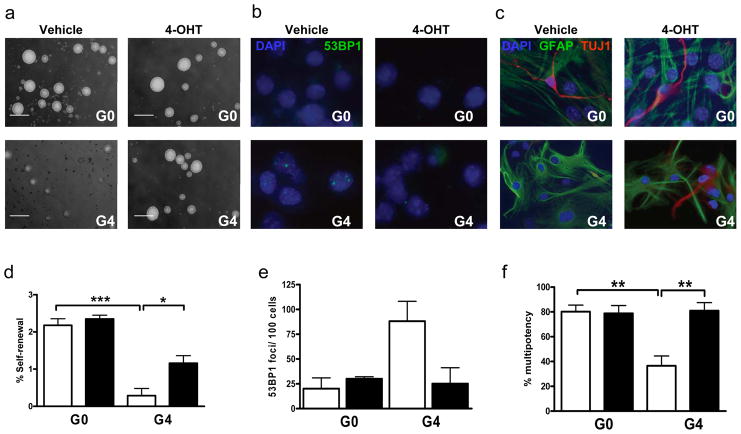
An ageing world population has fuelled interest in regenerative remedies that may stem declining organ function and maintain fitness. Unanswered is whether elimination of intrinsic instigators driving age-associated degeneration can reverse, as opposed to simply arrest, various afflictions of the aged. Such instigators include progressively damaged genomes. Telomerase-deficient mice have served as a model system to study the adverse cellular and organismal consequences of wide-spread endogenous DNA damage signalling activation in vivo. Telomere loss and uncapping provokes progressive tissue atrophy, stem cell depletion, organ system failure and impaired tissue injury responses. Here, we sought to determine whether entrenched multi-system degeneration in adult mice with severe telomere dysfunction can be halted or possibly reversed by reactivation of endogenous telomerase activity. To this end, we engineered a knock-in allele encoding a 4-hydroxytamoxifen (4-OHT)-inducible telomerase reverse transcriptase-oestrogen receptor (TERT-ER) under transcriptional control of the endogenous TERT promoter. Homozygous TERT-ER mice have short dysfunctional telomeres and sustain increased DNA damage signalling and classical degenerative phenotypes upon successive generational matings and advancing age. Telomerase reactivation in such late generation TERT-ER mice extends telomeres, reduces DNA damage signalling and associated cellular checkpoint responses, allows resumption of proliferation in quiescent cultures, and eliminates degenerative phenotypes across multiple organs including testes, spleens and intestines. Notably, somatic telomerase reactivation reversed neurodegeneration with restoration of proliferating Sox2(+) neural progenitors, Dcx(+) newborn neurons, and Olig2(+) oligodendrocyte populations. Consistent with the integral role of subventricular zone neural progenitors in generation and maintenance of olfactory bulb interneurons, this wave of telomerase-dependent neurogenesis resulted in alleviation of hyposmia and recovery of innate olfactory avoidance responses. Accumulating evidence implicating telomere damage as a driver of age-associated organ decline and disease risk and the marked reversal of systemic degenerative phenotypes in adult mice observed here support the development of regenerative strategies designed to restore telomere integrity.


Conflict of interest statement

The authors declare no competing financial interests.

Figures

Figure 1
Figure 1. 4-OHT-dependent induction of telomerase activity in TERT-ER cells
(a, top) Telomerase activity in eNSCs (* telomerase products); (a, bottom) real-time quantitation of reactions above; (b) representative G4TERT-ER splenocyte metaphases; (c) proliferation of adult G4TERT-ER fibroblasts (n=3) in media with vehicle (black) or 4-OHT (red); (d) representative image of G4TERT-ER fibroblasts (passage 6) in media with 4-OHT (bottom) or vehicle (top) ; (e) signal-free ends in primary splenocyte metaphases, 15 metaphases/sample, n=2 (*p<0.05); (f) mean telomere-FISH signal in primary splenocyte interphases, normalized to centromeric signal, n=3 (***p<0.0001). Open bars correspond to vehicle-treated and filled bars to 4-OHT-treated, error bars represent s.d.
Figure 2
Figure 2. Telomerase activation in adult TERT-ER mice
Representative tissue images from age-matched experimental and control mice (a) H&E stained sections of testes; (b) primary splenocytes stained for 53BP1; (c) small intestine sections stained for 53BP1; (d) testes weight of adult males (30–50 w.o., n≥10); (e) 53BP1 nuclear foci/100 nuclei (n=3); (f) 53BP1 nuclear foci/100 crypts (n=4†); (g) litter sizes (n=3); (h) spleen weights (n≥6); (i) apoptotic (TUNEL+) cells/100 intestinal crypts (n≥20). ***p=0.0001, **p<0.005, *p<0.05. Open bars correspond to vehicle-treated and filled bars to 4-OHT-treated groups, error bars represent s.d.
Figure 3
Figure 3. Neural stem cell function following telomerase reactivation in vitro
Representative images of experimental and control mice-derived (a) neurospheres, (b) differentiated NSCs stained with 53BP1 or (c) GFAP and TUJ1 antibodies. (d) Self-renewal capacity of secondary neurospheres (n=4) ***p<0.0001, *p<0.001. (e) Percentage of cells displaying 53BP1 nuclear foci (>400 nuclei/culture, n=3). (f) Multipotency (GFAP+/TUJ1+) of NSCs (n=4, 308 wells/culture condition) **p=0.0066. Scale bar represents 100μm. Open bars correspond to vehicle-treated and filled bars to 4-OHT-treated groups, error bars represent s.d.
Figure 4
Figure 4. NSC proliferation and differentiation following telomerase reactivation in vivo
NSC proliferation and neurogenesis were measured by Ki67, Sox-2 and DCX expression in SVZ from experimental and control mice. Mature oligodendrocytes in the CC were stained with Olig2. Equivalent coronal sections (n>10) were scored in a blinded fashion by laser scanning and plotted on the right panels. 20X (SVZ) or 40X (CC) objectives were used. *** p<0.0001, **p=0.0022. Open bars correspond to vehicle-treated and filled bars to 4-OHT-treated groups, error bars represent s.d.
Figure 5
Figure 5. Myelination, brain size, and olfactory function following telomerase reactivation
(a) Representative brains from age-matched experimental and control animals; (b) brain weights, n≥10, ***p=0.0004, *p=0.02; (c) representative electron micrographs of myelinated axonal tracts in CC; arrow heads indicate myelin sheath width (12,000X); scale bars=200nm; (d) g ratios (inner/outer radii) (n=2, >150 axons/mouse) ***p<0.0001; (e) representative tracings of experimental and control mice during 3-minute exposure to water or 2-MB. (f, g) Time spent in scent zone with water or 2-MB for vehicle- or 4-OHT-treated G0TERT-ER (squares) and G4TERT-ER (circles) mice; n=4. Error bars represent s.d., except in (d)(s.e.m.).

Comment in

  • Aging by telomere loss can be reversed.
    Bernardes de Jesus B, Blasco MA. Bernardes de Jesus B, et al. Cell Stem Cell. 2011 Jan 7;8(1):3-4. doi: 10.1016/j.stem.2010.12.013. Cell Stem Cell. 2011. PMID: 21211774

References

    1. Sahin E, Depinho RA. Linking functional decline of telomeres, mitochondria and stem cells during ageing. Nature. 7288;464:520–528. - PMC - PubMed
    1. Whitman MC, Greer CA. Adult neurogenesis and the olfactory system. Progress in neurobiology. 2009;89(2):162–175. - PMC - PubMed
    1. Sharpless NE, DePinho RA. How stem cells age and why this makes us grow old. Nature reviews. 2007;8(9):703–713. - PubMed
    1. Ju Z, Lenhard Rudolph K. Telomere dysfunction and stem cell ageing. Biochimie. 2008;90(1):24–32. - PubMed
    1. Hoeijmakers JH. DNA damage, aging, and cancer. The New England journal of medicine. 2009;361(15):1475–1485. - PubMed

Publication types

MeSH terms