. 2010 Dec;17(12):1414-21.
doi: 10.1038/nsmb.1961. Epub 2010 Nov 28.

The histone methyltransferase MLL1 permits the oscillation of circadian gene expression

Affiliations

The histone methyltransferase MLL1 permits the oscillation of circadian gene expression

Sayako Katada et al. Nat Struct Mol Biol. 2010 Dec.

Abstract

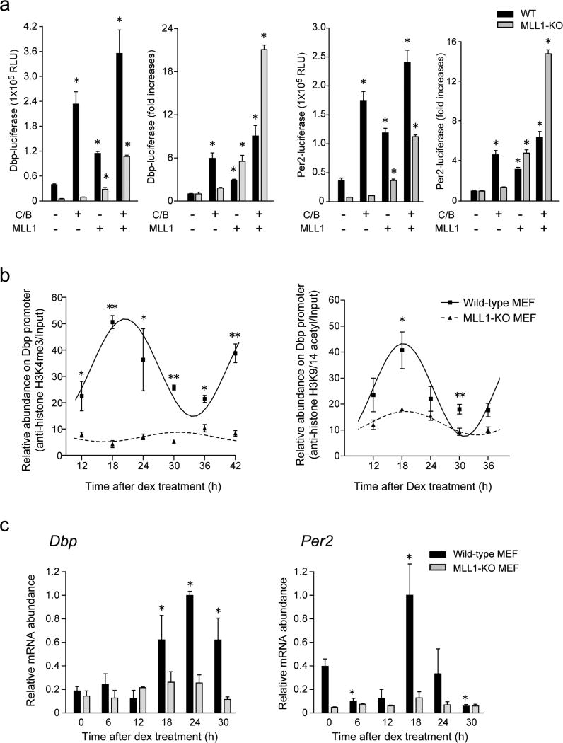
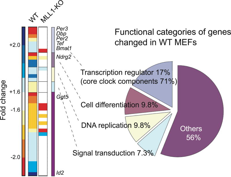
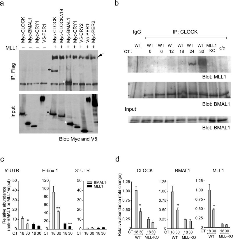
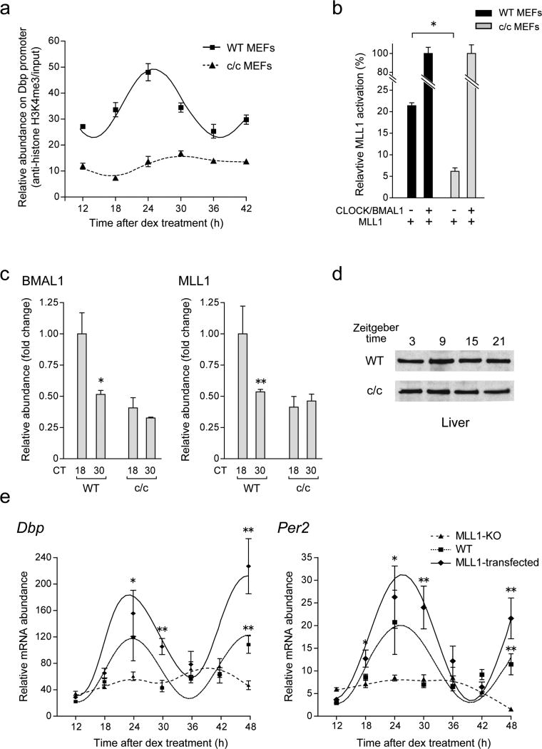
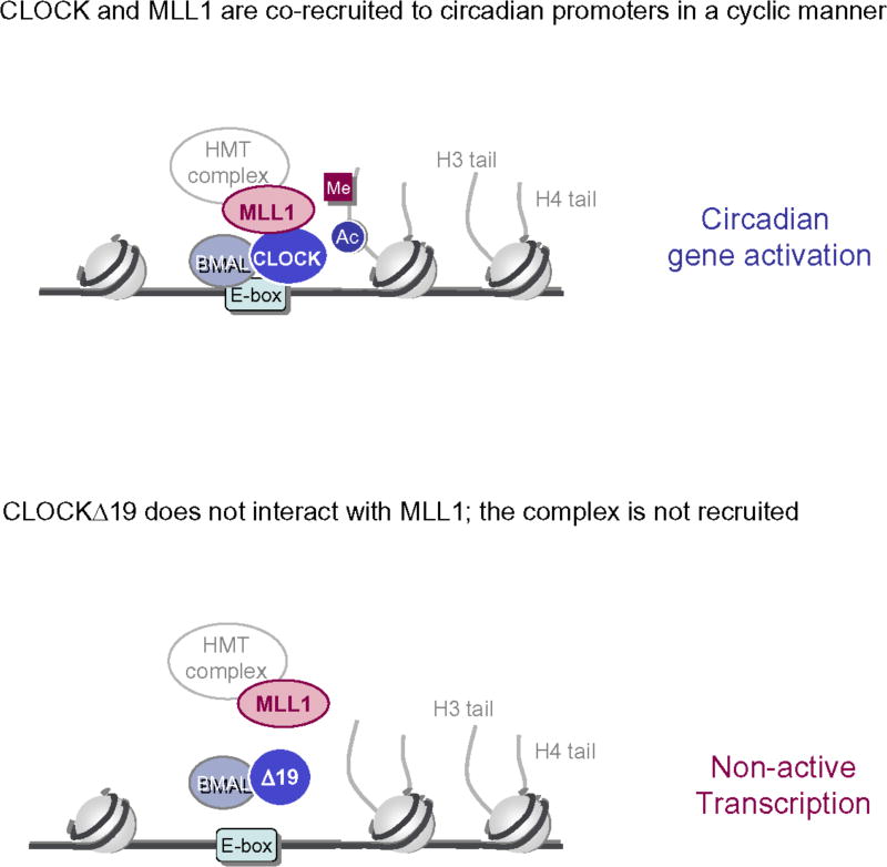
The classical view of the molecular clock is based on interlocked transcriptional-translational feedback loops. Because a substantial fraction of the mammalian genome is expressed in a circadian manner, chromatin remodeling has been proposed to be crucial in clock function. Here we show that Lys4 (K4) trimethylation of histone H3 is rhythmic and follows the same profile as previously described H3 acetylation on circadian promoters. MLL1, a mammalian homolog of Drosophila trithorax, is an H3K4-specific methyltransferase implicated in transcriptional control. We demonstrate that MLL1 is essential for circadian transcription and cyclic H3K4 trimethylation. MLL1 is in a complex with CLOCK-BMAL1 and contributes to its rhythmic recruitment to circadian promoters and to H3 acetylation. Yet MLL1 fails to interact with CLOCKΔ19, providing an explanation for this mutation's dominant negative phenotype. Our results favor a scenario in which H3K4 trimethylation by MLL1 is required to establish a permissive chromatin state for circadian transcription.


Figures

Figure 1
Figure 1. Histone H3K4 methyltransferase MLL1 synergistically activate CLOCK:BMAL1-mediated gene transcription
(a) Me3 but not me1/2 level of H3K4 changes between two time points on the Dbp promoter E-box region. ChIP analyses were performed in MEFs at 18 or 30 hr after dexamethasone synchronization using antibodies against acetylated H3K9 and K14 (white) and me1, me2 or me3 H3K4 (black). (Means ±SEM of three independent samples, *p<0.05, and ***p<0.0001). The amount of highest modification time point (18 hr) was set to 1. (b) H3K4me3 follows same circadian rhythmicity as acetylation of H3K9/K14 on the Dbp promoter E-box region. ChIP analyses were performed in MEFs after dexamethasone synchronization. (Means ±SEM of three independent samples from a representative experiment. Analogous results were observed in three independent experiments). (c) Histone H3K4 specific-methyltransferase MLL1 and SET1A were transiently transfected with or without CLOCK:BMAL1 in 293 cells. MLL1 but not SET1A showed substantial increases of the dbp-luc transcription. On the other hand, methyltransferase-dead mutant of MLL1 (MLL1Δ)) abolished transcriptional activity. (Means ±SEM of eight samples in two independent experiments) (d) Synergistic transcriptional activation on E-box motif by MLL1 together with CLOCK:BMAL1. E-box motif-containing sequences (E-box), NF-κB-responsive element (NF-κB), estrogen responsive element (ERE) or PPARresponsive element (PPRE) luciferase reporter genes were transfected in presence or in absence of CLOCK:BMAL1 in 293 cells. (Means ±SEM of eight samples from two independent experiments).
Figure 2
Figure 2. MLL1 governs circadian oscillation of histone modifications and gene expression
(a) MLL1 deficient MEFs demonstrate impaired CLOCK:BMAL1 mediated transcriptional activities. MLL1 was expressed with or without CLOCK:BMAL1 in MEFs derived from wild type (black) or MLL1-KO (gray) mice. In MLL1-KO MEFs, CLOCK:BMAL1-mediated Dbp and Per2 promoter transactivation was basically abolished, however ectopic expression of MLL1 rescued the expression. (Means ±SEM of seven samples from two independent experiments, *p<0.001). (b) Circadian H3K4me3 is completely impaired in MLL1-KO MEFs. Dexamethasone treated wild type (square) and MLL1-KO (triangle) MEFs were analyzed by ChIP using anti-H3K4me3 (left) or anti-acetylated H3K9/14 antibodies (right). (Means ±SEM of three independent samples, ANOVA p<0.001, and *p<0.05, **p<0.001 at t-test) (c) Circadian rhythmic expression of Dbp and Per2 were dramatically decreased in MLL1-KO MEFs. mRNA was extracted from wild type (black) or MLL1-KO (gray) MEFs after dexamethasone shock and analyzed by quantitative real-time PCR. The amount of highest expression time point was set to 1. (Means ±SEM of three samples, ANOVA p<0.001, and *p<0.05 at t-test)
Figure 3
Figure 3. Global analysis of gene expression profile in MLL1-KO MEFs
mRNA was extracted from wild type or MLL1-KO MEFs post 18hr or 30hr after serum shock, and microarray analyses were performed using GeneChip Mouse Gene 1.0 ST Array. Forty-one genes out of 35557 probes were identified as genes whose expression levels were significantly changed (>1.6 fold) between two-time point in WT MEFs. Heat map represent the fold changes of 41 genes in WT and MLL1-KO MEFs. Pie chart represent functional categories of genes changing in wild type MEFs. Eight out of 41 gene were listed as typical circadian regulated genes.
Figure 4
Figure 4. MLL1 interacts with CLOCK and BMAL1 and recruited to chromatin in circadian fashion
(a) CLOCK and BMAL1, but not CLOCKΔ19, were co-immunoprecipitated with MLL1. 293 cells were transfected with indicated expression vectors together with or without Flag-MLL1-Myc. Flag-tagged MLL1 N-terminal fragments (320 kDa) were immunoprecipitated by Flag-Agar, and coimmunoprecipitated proteins were determined by western analysis using anti-Myc and anti-V5 antibodies. Together with Myc-tagged 180 kDa MLL1 C-terminal fragments (arrow), Myc-BMAL1 (white asterisk) and Myc-CLOCK (black asterisk) proteins were coimmunoprecipitated with MLL1 N-terminal fragments. On the other hand, CLOCKΔ19, CRY1, CRY2, PER1 and PER2 were not co-immunoprecipitated with MLL1. Lower panel shows the results of total cell lysates as an input. (b) Endogenous MLL1 interacts with CLOCK in a circadian manner. MEFs were entrained by serum shock and harvested at various circadian times, then cellular extracts were immunoprecipitated using anti-CLOCK antibody. Co-immunoprecipitated proteins were determined using anti-BMAL1 and anti-MLL1 antibodies. Lower panel shows the results of total cell lysates as an input. (c) MLL1 recruited to E-box proximal region of the Dbp promoter in a circadian manner. ChIP analyses were performed on dual crosslinked MEFs at 18 or 30 hr after dexamethasone synchronization using antibodies against BMAL1 (gray) and MLL1 (black), and quantitative PCR was performed using 5’-UTR, E-box, or 3’-UTR primers. (Means ±SEM of six independent samples. *p<0.05, and **p<0.01) (d) ChIP analyses on the Dbp promoter E-box region in wild type or MLL1-KO MEFs. Analyses were performed on dual crosslinked MEFs at 18 or 30 hr after dexamethasone treatment using antibodies against BMAL1, CLOCK, and MLL1. The amount of highest binding time point (18 hr) in wild type MEFs was set to 1. (Means ±SEM of six independent samples. *p<0.01)
Figure 5
Figure 5. Impaired H3K4me3 and less recruitment of BMAL1 and MLL1 proteins in c/c mutant MEFs
(a) Rhythmic H3K4me3 was completely abolished in c/c mutant MEFs. ChIP analyses were performed on the Dbp promoter E-box region in wild-type (square) or c/c mutant (triangle) MEFs at each time point post dexamethasone shock. (b) Luciferase reporter gene assay using the Dbp promoter in c/c mutant MEFs. MLL1 was transiently transfected with or without CLOCK:BMAL1 in wild type (black) or c/c mutant (gray) MEFs, and luciferase activity was measured post 48 hr. Relative light units obtained from each CLOCK:BMAL1 and MLL1 transfected MEFs were set to 100 (%). (Means ±SEM of four independent samples. *p<0.001) (c) Circadian recruitment of BMAL1 and MLL1 on the Dbp promoter E-box regions were abolished in c/c mutant MEFs. Dexamethasone shocked and dual crosslinked wild-type and c/c mutant MEFs were analyzed by ChIP using anti-BMAL1 (left) or anti-MLL1 (right) antibodies. (Means ±SEM of three independent samples. *p<0.05, **p=0.0512) (d) MLL1 protein levels of MLL1 do not change in the liver from c/c mice. Proteins were extracted from each time point of liver from WT or c/c mutant mice and MLL1 expression levels were detected using anti-MLL antibodies. (e) Rescue of circadian mRNA expression of clock genes by stably expressing MLL1 in MLL1-KO MEFs. mRNA was extracted from wild type (square), Flag-MLL1 stably expressed MLL1-KO (diamond), or mock transfected MLL1-KO (triangle) MEFs after dexamethasone shock and Dbp and Per2 expression levels were analyzed by quantitative real-time PCR. (Means ±SEM of four independent samples. *p<0.05, **p<0.001)
Figure 6
Figure 6
CLOCK and MLL1 physically interact at the level of clock-controlled promoters (with E-boxes). Together induce PTMs on histone N-terminal tails associate to circadian transcriptional activation. The mutant protein CLOCKΔ19 fails to associate with MLL1 (lower figure), providing a molecular explanation of its impaired activation potential. As MLL1 and CLOCKΔ19 do not interact, there is also lack of PTMs on the H3 tail, leading to lack of circadian transcription.

References

    1. Dunlap JC, Loros JJ, Liu Y, Crosthwaite SK. Eukaryotic circadian systems: cycles in common. Genes Cells. 1999;4:1–10. - PubMed
    1. Reppert SM, Weaver DR. Coordination of circadian timing in mammals. Nature. 2002;418:935–41. - PubMed
    1. Bell-Pedersen D, et al. Circadian rhythms from multiple oscillators: lessons from diverse organisms. Nat Rev Genet. 2005;6:544–56. - PMC - PubMed
    1. Schibler U, Sassone-Corsi P. A web of circadian pacemakers. Cell. 2002;111:919–22. - PubMed
    1. Fu L, Lee CC. The circadian clock: pacemaker and tumour suppressor. Nat Rev Cancer. 2003;3:350–61. - PubMed

Publication types

MeSH terms