. 2010 Sep;1(5):329-38.
doi: 10.18632/oncotarget.165.

CD277 is a negative co-stimulatory molecule universally expressed by ovarian cancer microenvironmental cells

Affiliations

CD277 is a negative co-stimulatory molecule universally expressed by ovarian cancer microenvironmental cells

Juan R Cubillos-Ruiz et al. Oncotarget. 2010 Sep.

Abstract

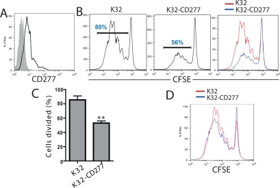
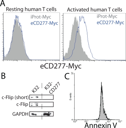
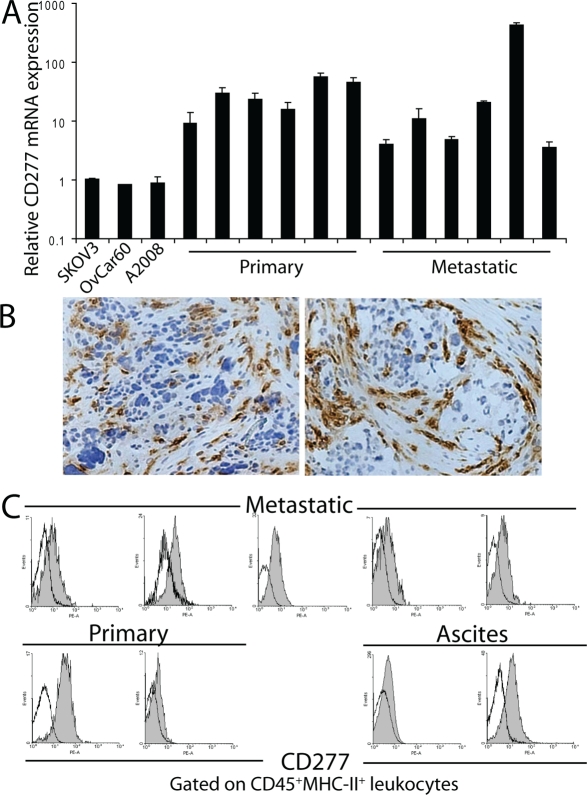
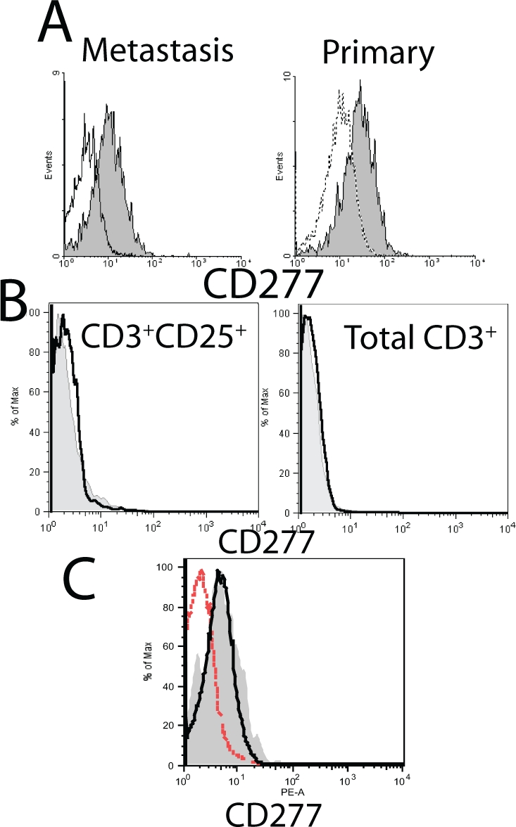
CD277, a member of the butyrophilin subfamily 3 (BTN3), shares significant sequence similarities and predicted common structural features with inhibitory B7-H4 and other members of the B7 superfamily. Here we report that CD277 is consistently expressed in stromal, as well as tumor cells in the microenvironment of human advanced ovarian carcinoma specimens, both of primary and metastatic origin. MHC-II+ myeloid antigenpresenting leukocytes (dendritic cells and macrophages) express significantly higher levels of surface CD277, compared to other tumor-infiltrating leukocyte subsets, and this expression is significantly up-regulated by multiple common tumor microenvironmental signals, including VEGF and CCL3. Most importantly, engagement of CD277 on the surface of TCR-stimulated T cells inhibits their otherwise robust expansion and production of Th1 cytokines by preventing the up-regulation of cFLIP. Our results point to a role for CD277 up-regulated by microenvironmental signals in the acquisition of a regulatory phenotype by tumor-associated myeloid cells. Consequently, CD277, and likely other butyrophilins and butyrophilin-like molecules, emerge as regular players in the orchestration of immunosuppressive networks in ovarian cancer, and therefore new targets for interventions to overcome immune evasion and boost anti-tumor immunity in cancer patients.

Keywords: Tumor microenvironment; butyrophilin; dendritic cell; immune evasion; immunotherapy; tumor immunology.


Conflict of interest statement

The authors declare no conflict of interest.

Figures

Figure 1:
Figure 1:. CD277 shares sequence and structural similarity with other members of the B7 superfamily of costimulatory molecules
(A) Alignment of CD277 and B7-H4. Conserved cysteine residues are highlighted in red. Conserved Amino Acid regions are highlighted in black. (B) Cluster analysis of B7 superfamily members. The Amino Acid sequence of CD277 was aligned to other B7 superfamily members and a similarity tree was generated. (C) (left and center) Presentation of the CD277 and B7-H4 structures, predicted using the know structure of PD-L1 as a model, based on sequence. (right) Schematic overlay of both predicted structures. Blue, CD277; Orange, B7-H4.
Figure 2:
Figure 2:. CD277 inhibits TCR-mediated human T cell proliferation
(A) FACS analysis of CD277 expression by mock-transduced K32 (aAPCs) cells (solid) and K32 cells stably infected with a retroviral construct encoding CD277 (open). (B) Untransduced or CD277-expressing K32 cells were loaded with anti-CD3 agonistic antibodies (OKT3; 100 ng/ml) and cells were then co-cultured (1:10) with CFSE-labeled human peripheral blood T cells. The percentage of proliferating T cells was analyzed by FACS after five days of proliferation. Data are representative of five independent experiments with similar results. (C) Representative FACS analysis of the data shown in B. **P<0.05 (Mann-Whitney). (D) CD28 costimulation alleviates CD277-mediated suppression. K32 cells were loaded with anti-CD3 and anti-CD28 agonistic antibodies (25 ng) and cells were cocultured with human T cells, as described above. CFSE dilution was evaluated by FACS five days later. Data are representative of four independent experiments with similar results
Figure 3:
Figure 3:. CD277 inhibits cFLIP in activated human T cells
(A) Resting human T cells or T cells activated for 24 hours with anti-CD3/anti-CD28 beads were incubated with a Myc-tagged irrelevant protein or a with Myc-tagged extracellular form of CD277. Protein binding to the cell surface was analyzed by FACS using a secondary anti-Myc antibody. Data are representative of three independent experiments with similar results. iProt, irrelevant protein. eCD277, extracellular region of CD277. (B) Human T cells were cocultured for 24 hours with K32 cells ectopically expressing or not CD277 and whole cell lysates were used to analyze c-Flip expression by Western blot. Data are representative of two independent experiments with similar results. (C) Resting human T cells were activated for 24 hours with K32 cells ectopically expressing (solid) or not (open) CD277 and intracellular expression of Annexin V was determined.
Figure 4:
Figure 4:. CD277 inhibits Th1 cytokine secretion by TCR-stimulated human T cells
K32 cells expressing or not CD277 were loaded with anti-CD3 agonistic antibodies and cells were cocultured with human T cells as described above. Culture supernatants were collected after five days and cytokine secretion was analyzed by Multiplex analysis. Data are representative of two independent experiments with similar results. **P<0.05 (Mann-Whitney).
Figure 5:
Figure 5:. CD277 is abundantly expressed in the microenvironment of human epithelial ovarian cancer
(A) Real Time RT-PCR analysis of CD277 mRNA levels in human ovarian cancer cells lines (SKOV3, Ovcar60 and A2008) and multiple human tumor specimens. (B) Representative immunohistochemistry of CD277 protein expression in ovarian tumors from two different patients (magnification, x200). (C) Single-cell suspensions from different stage III-IV human ovarian tumors and ascites were procured and surface expression of CD277 on CD45+MHC-II+ leukocytes (CD20-)[13] was determined by FACS. Open histograms represent staining with isotype control antibodies, solid histograms indicate CD277 staining.
Figure 6:
Figure 6:. CD277 is preferentially expressed by APCs and tumor cells in the ovarian carcinoma microenvironment
(A) Representative FACS analysis of CD277 expression on CD45+MHC-II+CD20- (solid), compared to that of other CD45+ cells (open) within the same human specimens. (B) FACS analysis of CD277 on CD4+CD25+ (regulatory + activated T cells) and total CD3+ T cells in a metastatic ovarian cancer tumor. Solid histograms indicate CD277 expression, open histograms represent isotype control staining. Identical results were obtained when primary tumors or ascites were analyzed for CD277 expression on T cells. (C) Representative FACS analysis of CD277 expression on CD45- cells in dissociated ovarian cancer specimens (mainly tumor cells; open thicked), compared to that in CD45+MHC-II+CD20- inflammatory leukocytes (solid) in the same specimen. Open dashed histograms represent isotype control staining. Comparable results were obtained in primary and metastatic tumors.
Figure 7:
Figure 7:. Tumor microenvironmental inflammatory cytokines and hypoxia-associated mediators up-regulate the expression of CD277 in monocyte-derived human DCs
Monocyte-derived DCs were incubated for 18 hours with the indicated cytokines and CD277 expression was evaluated by Real Time Quantitative PCR.

References

    1. Jemal A, Siegel R, Ward E, Hao Y, Xu J, Thun MJ. Cancer statistics. CA Cancer J Clin. 2009;59:225–49. 2009. - PubMed
    1. Jemal A, Siegel R, Ward E, Hao Y, Xu J, Murray T, Thun MJ. Cancer statistics. CA Cancer J Clin. 2008;58:71–96. 2008. - PubMed
    1. Zhang L, Conejo-Garcia JR, Katsaros D, Gimotty PA, Massobrio M, Regnani G, Makrigiannakis A, Gray H, Schlienger K, Liebman MN, Rubin SC, Coukos G. Intratumoral T cells, recurrence, and survival in epithelial ovarian cancer. N Engl J Med. 2003;348:203–13. - PubMed
    1. Coukos G, Conejo-Garcia JR, Roden RB, Wu TC. Immunotherapy for gynaecological malignancies. Expert Opin Biol Ther. 2005;5:1193–210. - PubMed
    1. Conejo-Garcia JR, Benencia F, Courreges MC, Gimotty PA, Khang E, Buckanovich RJ, Frauwirth KA, Zhang L, Katsaros D, Thompson CB, Levine B, Coukos G. Ovarian carcinoma expresses the NKG2D ligand Letal and promotes the survival and expansion of CD28- antitumor T cells. Cancer Res. 2004;64:2175–182. - PubMed

Publication types

MeSH terms