Review
. 2011 Aug;26(8):1181-95.
doi: 10.1007/s00467-010-1697-5. Epub 2010 Nov 27.

Cystic diseases of the kidney: ciliary dysfunction and cystogenic mechanisms

Affiliations
Review

Cystic diseases of the kidney: ciliary dysfunction and cystogenic mechanisms

Cecilia Gascue et al. Pediatr Nephrol. 2011 Aug.

Abstract

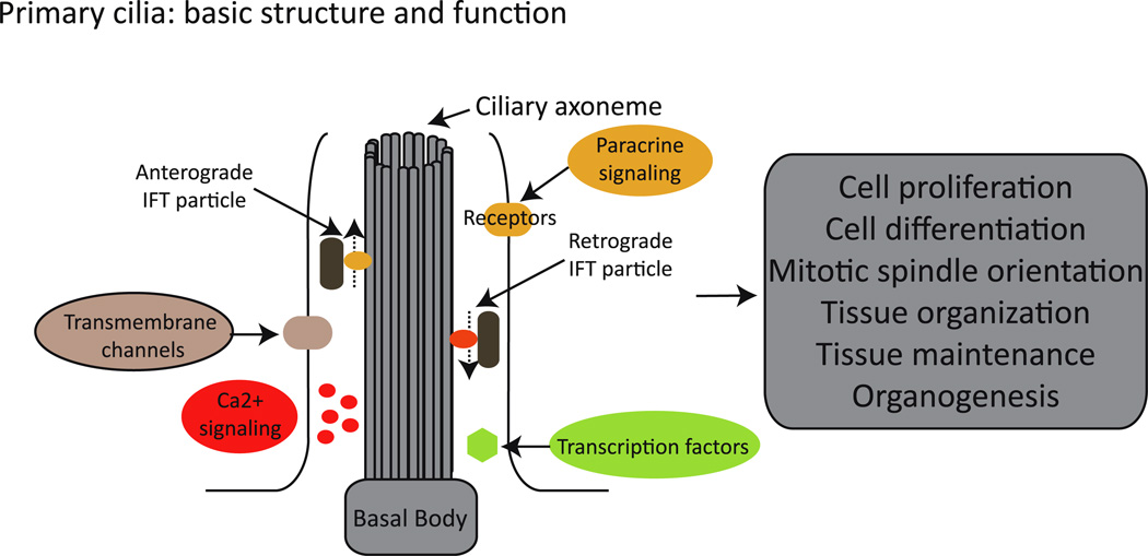
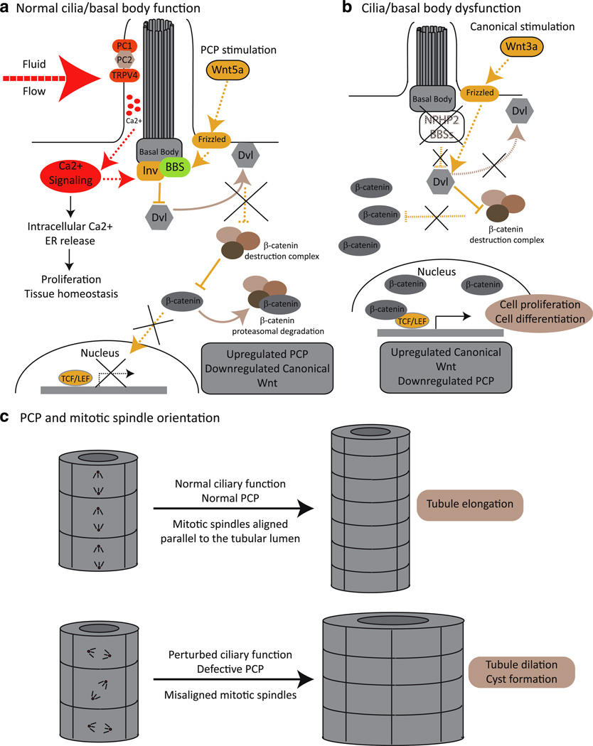
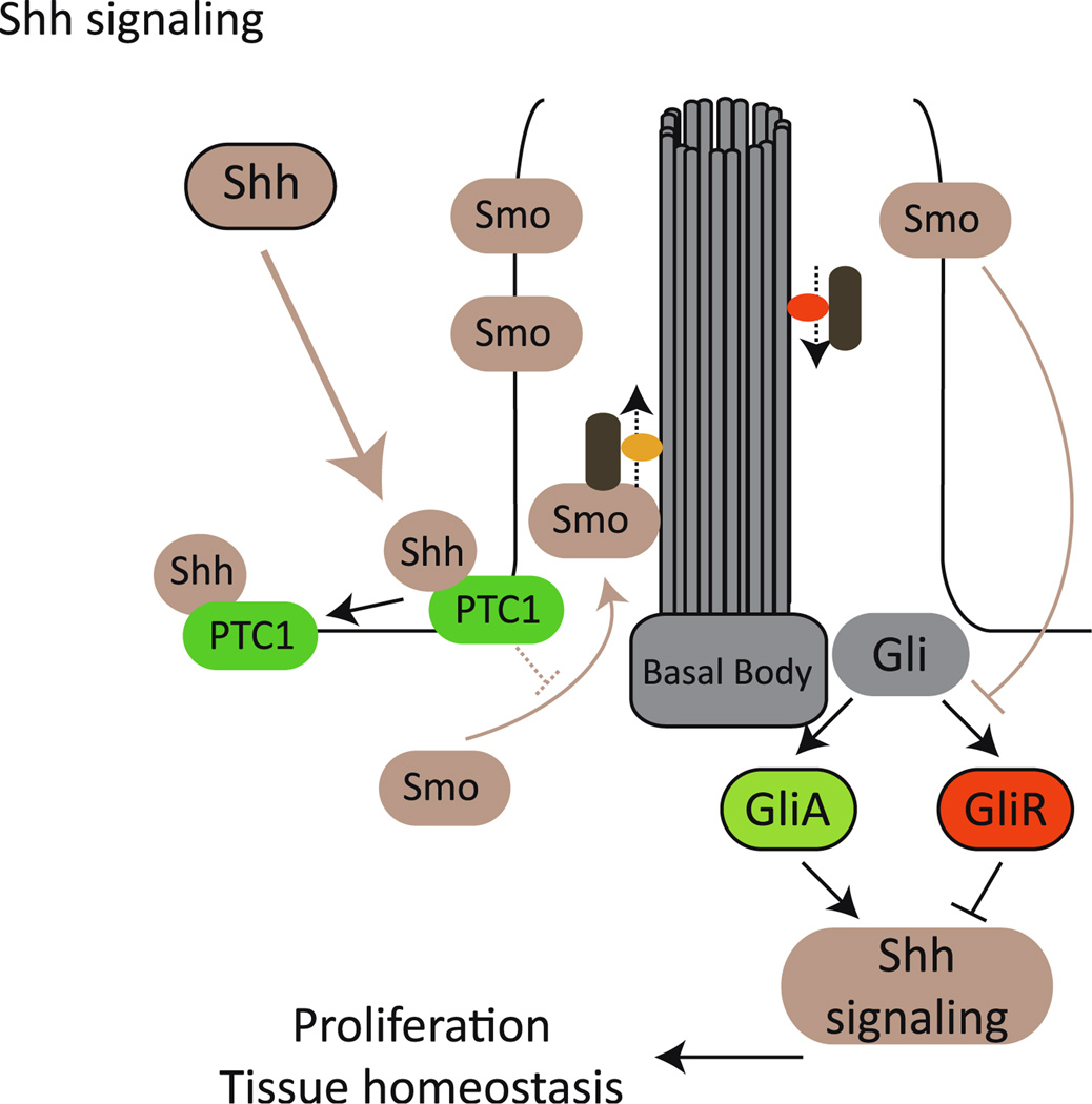
Ciliary dysfunction has emerged as a common factor underlying the pathogenesis of both syndromic and isolated kidney cystic disease, an observation that has contributed to the unification of human genetic disorders of the cilium, the ciliopathies. Such grouping is underscored by two major observations: the fact that genes encoding ciliary proteins can contribute causal and modifying mutations across several clinically discrete ciliopathies, and the emerging realization that an understanding of the clinical pathology of one ciliopathy can provide valuable insight into the pathomechanism of renal cyst formation elsewhere in the ciliopathy spectrum. In this review, we discuss and attempt to stratify the different lines of proposed cilia-driven mechanisms for cystogenesis, ranging from mechano- and chemo-sensation, to cell shape and polarization, to the transduction of a variety of signaling cascades. We evaluate both common trends and differences across the models and discuss how each proposed mechanism can contribute to the development of novel therapeutic paradigms.


Figures

Fig. 1
Fig. 1
The cilium: basic structure and function. Cilia are formed by a microtubule core (axoneme) that is organized from a basal body. Along the axoneme, intraflagellar transport (IFT) particles are transported in (anterograde) and out (retrograde) of the cilium. The cilium concentrates and organizes a number of channels, receptors, and effectors, therefore playing a critical role in, for example, Ca2+ and paracrine signaling, ultimately regulating cellular, tissue, and organ homeostasis
Fig. 2
Fig. 2
Primary cilia: mechano-sensors and Wnt transducers. a PC1, PC2, and TRPV4 form a Ca2+ channel that localizes to the primary cilium and which upon a mechanical stimulus, such as fluid flow, initiate a Ca2+ signaling cascade that regulates cell proliferation. Functional cilia are also needed to maintain the tight balance between canonical and noncanonical [planar cell polarity (PCP)] Wnt signaling. In the presence of a functional cilium, and upon PCP stimulation and Ca2+ signaling, basal bodies proteins, such as the BBSs and inversin (Inv)/NPHP2, mediate the degradation and/or relocalization of Disheveled (Dvl) to the cellular membrane. In this scenario, Dvl is no longer able to suppress the β-catenin destruction complex, and β-catenin is therefore degraded by the proteasome and is unable to act as a transcriptional activator. b When cilia and/or basal body function is compromised, canonical Wnt signals (for example, Wnt 3a) are not antagonized, Dvl is able to repress the β-catenin destruction complex, and β-catenin accumulates in the nucleus and drives the expression of TCF–LEF responsive genes. c The non-canonical PCP Wnt pathway is required for providing positional information to cells allowing the correct alignment of their mitotic spindles with respect to the lumen of the renal tubule. Upon ciliary dysfunction, the PCP signal is defective, and cell division orients randomly. Therefore, under normal ciliary function, cell division in the renal tubules results in tubular elongation, while ciliary dysfunction results in tubule dilation, facilitating cystogenesis
Fig. 3
Fig. 3
Primary cilia and sonic hedgehog (Shh) transduction. Binding of Shh to Patched 1 receptor (PTC1) results in the re-localization of Smoothened (Smo) into the ciliary compartment, thus inhibiting the formation of GliR (repressor) and favoring the processing of Gli into GliA, the activator that drives the expression of different target genes

References

    1. Beales PL, Parfrey PS, Katsanis N. The Bardet-Biedl and Alstrom Syndromes. In: Maher E, Saggar-Malik A, editors. Genetics of renal disease. Oxford: Oxford University Press; 2004. pp. 361–398.
    1. Hildebrandt F, Otto E. Cilia and centrosomes: a unifying pathogenic concept for cystic kidney disease? Nat Rev Genet. 2005;6:928–940. - PubMed
    1. Watnick T, Germino G. From cilia to cyst. Nat Genet. 2003;34:355–356. - PubMed
    1. Badano JL, Mitsuma N, Beales PL, Katsanis N. The ciliopathies: an emergin class of human genetic disorders. Annu Rev Genomics Hum Genet. 2006;7:125–148. - PubMed
    1. Fliegauf M, Benzing T, Omran H. When cilia go bad: cilia defects and ciliopathies. Nat Rev Mol Cell Biol. 2007;8:880–893. - PubMed

Publication types

MeSH terms