. 2011 Jul;21(4):428-40.
doi: 10.1111/j.1750-3639.2010.00466.x. Epub 2011 Jan 10.

Axonal loss and neurofilament phosphorylation changes accompany lesion development and clinical progression in multiple sclerosis

Affiliations

Axonal loss and neurofilament phosphorylation changes accompany lesion development and clinical progression in multiple sclerosis

Lucas Schirmer et al. Brain Pathol. 2011 Jul.

Abstract

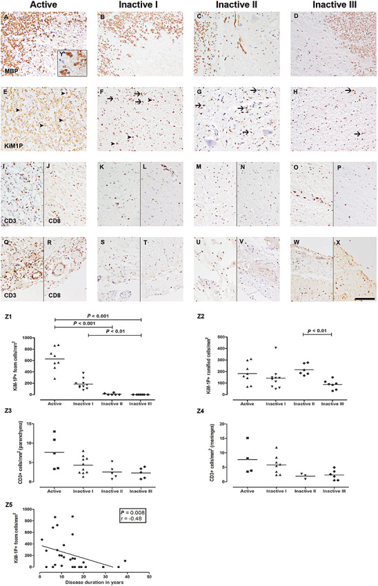
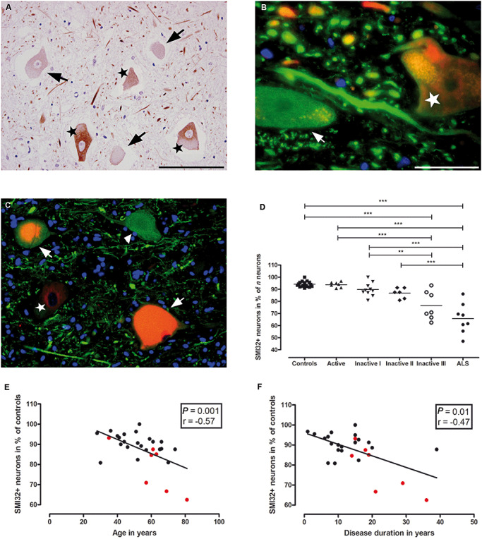
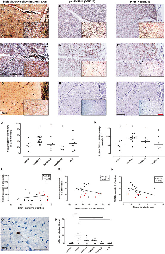
Neuroaxonal damage and loss are increasingly recognized as disability determining features in multiple sclerosis (MS) pathology. However, little is known about the long-term sequelae of inflammatory demyelination on neurons and axons. Spinal cord tissue of 31 MS patients was compared to three amyotrophic lateral sclerosis (ALS) and 10 control subjects. MS lesions were staged according to the density of KiM-1P positive macrophages and microglia and the presence of myelin basic protein (MBP) positive phagocytes. T cells were quantified in the parenchyma and meninges. Neuroaxonal changes were studied by immunoreactivity (IR) for amyloid precursor protein (APP) and variably phosphorylated neurofilaments (SMI312, SMI31, SMI32). Little T cell infiltration was still evident in chronic inactive lesions. The loss of SMI32 IR in ventral horn neurons correlated with MS lesion development and disease progression. Similarly, axonal loss in white matter (WM) lesions correlated with disease duration. A selective reduction of axonal phosphorylated neurofilaments (SMI31) was observed in WM lesions. In ALS, the loss of neuronal SMI32 IR was even more pronounced, whereas the relative axonal reduction resembled that found in MS. Progressive neuroaxonal neurofilament alterations in the context of chronic inflammatory demyelination may reflect changes in neuroaxonal metabolism and result in chronic neuroaxonal dysfunction as a putative substrate of clinical progression.


Figures

Figure 1
Figure 1
Staging of spinal multiple sclerosis (MS) lesions and T cell pathology. Spinal MS lesions were staged according to the presence of MBP+ (A–D) myelin degradation products in phagocytes (Active, A, E) and the density of KiM‐1P+ (E–H) macrophages/microglia (Inactive I–III, B–D, F–H), identified by morphological criteria: KiM‐1P immunohistochemistry revealed foam cells of variable density in Active and Inactive I lesions (E, F, arrowheads) and ramified cells of microglia phenotype in all lesion stages (E–H, arrows). Actively demyelinating lesions (Active) showed foamy phagocytes with incorporated MBP+ myelin degradation products (A and insert Y). Lesion stages Inactive I–III designate inactive demyelinated lesions with variable numbers of KiM‐1P+ cells (F–H) and a loss of MBP+ myelin sheaths (B–D). Few foam cells were still present in Inactive I lesions (B). Stage Inactive III harbored the lowest density of KiM‐1P+ cells (H). Foam cell densities decreased from lesion stages Active to Inactive I and Inactive II/III (Z1). Ramified microglia cell densities were significantly lower in lesions typed Inactive III compared to Inactive II (Z2). Decrease of macrophage like KiM‐1P+ cells correlated with the disease duration of the patients (Z5). Highest numbers of parenchymal and meningeal CD3+ and CD8+ T cells were found in stages Active (I, J and Q, R) and Inactive I (K, L and S, T). Sections with Inactive II (M, N and U, V) and Inactive III (O, P and W, X) lesions showed the lowest numbers of CD3+ and CD8+ cells. However, no statistically significant differences regarding CD3+ T cells in parenchymal lesions (Z3) and meninges (Z4) were observed. Scale bar (X): A–X: 100 µm; original magnifications: A–X: x200, Y: x1000.
Figure 2
Figure 2
Relative reduction of SMI32+ ventral spinal neurons in MS. SMI32 IR (nonP‐NF‐H) is observed in 94.2 ± 2.3% (mean ± SD) of ventral spinal neurons in control subjects (D). However, the percentage of SMI32+ neurons (A–C: marked with stars) was significantly lower in ventral horns of sections harboring demyelinated lesions with low densities of KiM‐1P positive cells and no foam cells, presumably late‐stage lesions [A (Inactive III): SMI32+ neurons = stars, SMI32− neurons = arrows; D (**P < 0.01, ***P < 0.001)]. In B (Inactive I) and C (Active), SMI32 IR (red, stars) in neuronal cell bodies is displayed adjacent to beta‐Tubulin+ (green, arrow B) and SMI35+[green, arrowhead (hypoP‐NF‐H), C] cell bodies with absent IR for SMI32. Of note, in C, an actively demyelinating MS lesion, chromatolytic ventral horn neurons (arrows) with double IR for SMI32 (red) and SMI35 (green) are seen. The decline in the percentage of SMI32+ neurons correlated with age at death of the patients (E; P = 0.0011, r = −0.5734) and disease duration (F; P = 0.0056, r = −0.5094). In E and F, spinal cord cross sections with lesions staged Inactive III (red circles) are indicated. Of note, active lesions occur predominantly in patients with shorter disease duration, whereas late‐stage inactive lesions are more frequent in patients with longer disease duration. Scale bars: A, C: 100 µm; B: 20 µm; original magnifications: A, C: x400; B: x1000.
Figure 3
Figure 3
Decline of axonal densities and neurofilament phosphorylation changes in axons. Axonal densities in control (A–C), MS (Inactive III; D–F) and amyotrophic lateral sclerosis (ALS) spinal cord white matter (G–I). Axon counts were carried out on sections stained with Bielschowsky silver impregnation (A, D, G) and antibodies against panP‐NF‐H (SMI312; B, E, H) and P‐NF‐H (SMI31; C, F, I). Lowest densities of Bielschowsky+ axons were found in Inactive III lesions (D, J). Low axonal densities in Active‐staged WM lesions were predominantly due to edema and inflammatory cell infiltration [J (**P < 0.01)]. The ratio SMI31+/Bielschowsky+ revealed an overproportional loss of SMI31+ axons in relation to Bielschowsky+ axons with lowest numbers in stages Active and Inactive III[D, F, K (*P < 0.05)]. The decrease of axonal phosphorylated neurofilaments (SMI31) was not paralleled by an increase in nonphosphorylated neurofilaments (SMI32; L). Similarly, no correlation was observed between the relative loss of neuronal SMI32 IR and the loss of n axons (Bielschowsky+) in spinal cord cross sections with inactive lesions (M). However, in inactive WM lesions, the relative loss of axonal SMI312 IR was correlated with increasing disease duration (N). In L–N, spinal cord cross sections with lesions staged Inactive III (red squares) are indicated. APP+ axonal spheroids (O) were predominantly found in Active WM lesions in MS (P) compared to controls and ALS (*P < 0.05; **P < 0.01; ***P < 0.001). Scale bars: A–I (insets): 200 µm (50 µm); original magnifications: A–I: x100 (x400).

References

    1. Agosta F, Absinta M, Sormani MP, Ghezzi A, Bertolotto A, Montanari E et al (2007) In vivo assessment of cervical cord damage in MS patients: a longitudinal diffusion tensor MRI study. Brain 130:2211–2219. - PubMed
    1. Bannerman PG, Hahn A, Ramirez S, Morley M, Bonnemann C, Yu S et al (2005) Motor neuron pathology in experimental autoimmune encephalomyelitis: studies in THY1‐YFP transgenic mice. Brain 128:1877–1886. - PubMed
    1. Barnett MH, Henderson AP, Prineas JW (2006) The macrophage in MS: just a scavenger after all? Pathology and pathogenesis of the acute MS lesion. Mult Scler 12:121–132. - PubMed
    1. Bitsch A, Schuchardt J, Bunkowski S, Kuhlmann T, Bruck W (2000) Acute axonal injury in multiple sclerosis. Correlation with demyelination and inflammation. Brain 123(Pt 6):1174–1183. - PubMed
    1. Bjartmar C, Kidd G, Mork S, Rudick R, Trapp BD (2000) Neurological disability correlates with spinal cord axonal loss and reduced N‐acetyl aspartate in chronic multiple sclerosis patients. Ann Neurol 48:893–901. - PubMed

Publication types

MeSH terms

Substances