. 2010 Nov;14(11):2687-92.
doi: 10.1111/j.1582-4934.2010.01185.x.

Cardiac telocytes: serial dynamic images in cell culture

Affiliations

Cardiac telocytes: serial dynamic images in cell culture

L Suciu et al. J Cell Mol Med. 2010 Nov.

Abstract

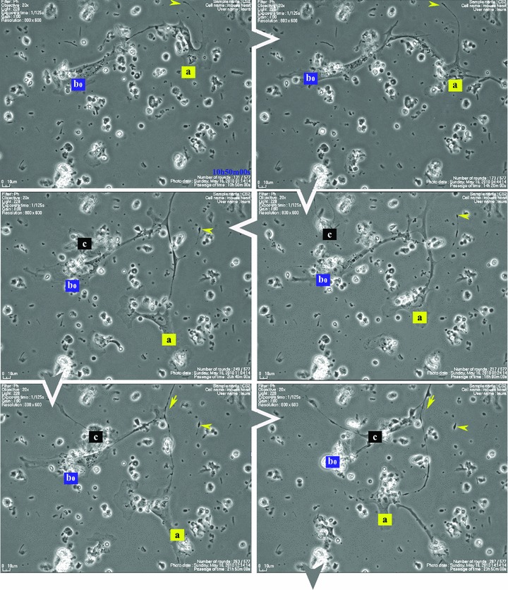
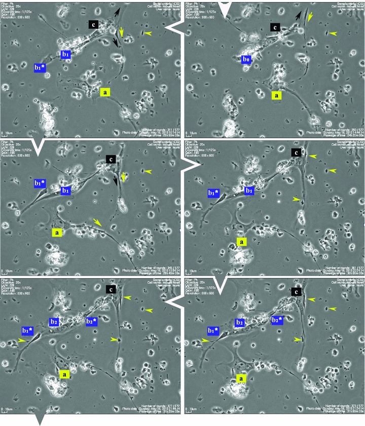
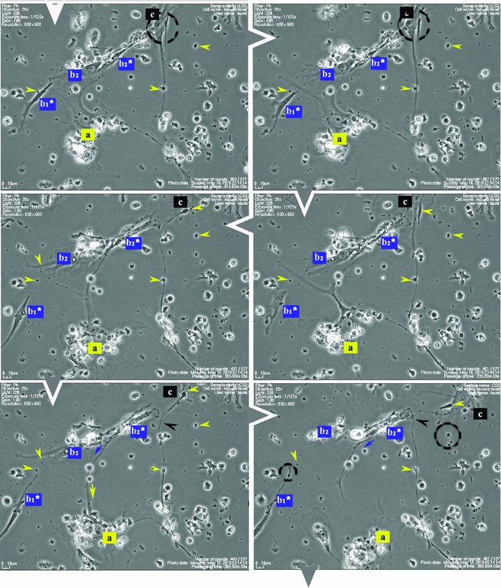
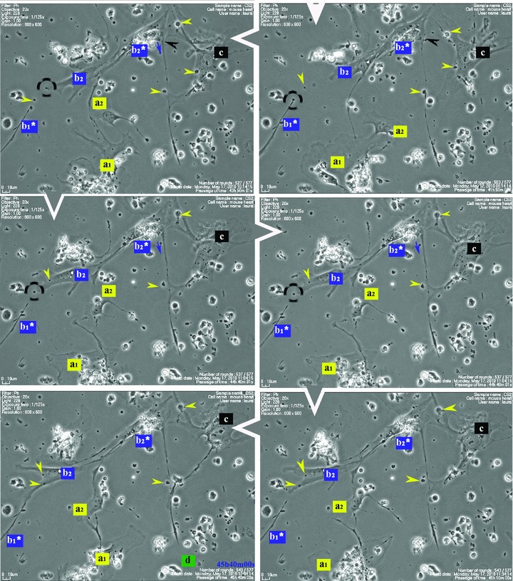
Telocytes (TC) are interstitial cells with telopodes (Tp). These prolongations (Tp) are quite unique: very long (several tens of micrometres) and very thin (≤0.5 μm), with moniliform aspect: thin segments (podomeres) alternating with dilations (podoms). To avoid any confusion, TC were previously named interstitial Cajal-like cells (ICLC). Myocardial TC were repeatedly documented by electron microscopy, immunohistochemistry and immunofluorescence. TC form a network by their Tp, either in situ or in vitro. Cardiac TC are (completely) different of 'classic' fibroblasts or fibrocytes. We hereby present a synopsis of monitoring, by time-lapse videomicroscopy, of Tp network development in cell culture. We used a protocol that favoured interstitial cell selection from adult mouse myocardium. Videomicroscopy showed dynamic interactions of neighbour TC during the network formation. During their movement, TC leave behind distal segments (podomeres) of their Tp as guiding marks for the neighbouring cells to follow during network rearrangement.


Figures

Fig 1
Fig 1
Serial snapshots selected from a time-lapse videomicroscopy recording of adult mouse myocardial interstitial cell culture. The chronological order of sequences is indicated by white arrowheads at the edge of each frame. TC were designated a, b, c and d in order of appearance in the microscopic field. They start structuring a network. The a TC (yellow) is the most active cell, scouting the area and planting marks (yellow arrowheads) out of its own regressing Tp (yellow arrows), for the other cells to follow. TC b2* and c are using those retracting Tp as guiding wires to grow their own cell projections (black and blue arrows). Following marks left by TC a (enclosed areas and yellow arrowheads), TC c leaves an entire Tp (black arrowhead) as a guiding mark for b2* and upcoming d cell. Note: a and b cells undergo cell division; daughter cells were assigned numeric indices.
Fig 1
Fig 1
Serial snapshots selected from a time-lapse videomicroscopy recording of adult mouse myocardial interstitial cell culture. The chronological order of sequences is indicated by white arrowheads at the edge of each frame. TC were designated a, b, c and d in order of appearance in the microscopic field. They start structuring a network. The a TC (yellow) is the most active cell, scouting the area and planting marks (yellow arrowheads) out of its own regressing Tp (yellow arrows), for the other cells to follow. TC b2* and c are using those retracting Tp as guiding wires to grow their own cell projections (black and blue arrows). Following marks left by TC a (enclosed areas and yellow arrowheads), TC c leaves an entire Tp (black arrowhead) as a guiding mark for b2* and upcoming d cell. Note: a and b cells undergo cell division; daughter cells were assigned numeric indices.

References

    1. Popescu LM, Faussone-Pellegrini MS. Telocytes – a case of serendipity: the winding way from interstitial cells of Cajal (ICC), via interstitial Cajal-like cells (ICLC) to Telocytes. J Cell Mol Med. 14:729–40. - PMC - PubMed
    1. Suciu L, Popescu LM, Gherghiceanu M, et al. Telocytes in human term placenta: morphology and phenotype. Cells Tissues Organs. 2010;192:325–39. - PubMed
    1. Popescu LM, Gherghiceanu M, Kostin S. Telocytes and heart renewing. In: Wang P, Kuo C-H, Takeda N, Singal PK, et al., editors. Adaptation biology and medicine. New Delhi, India: Narosa Publishing House Pvt. Ltd; 2010. pp. 17–39.
    1. Kostin S. Myocardial telocytes: a specific new cellular entity. J Cell Mol Med. 2010;14:1917–21. - PMC - PubMed
    1. Faussone-Pellegrini MS, Bani D. Relationships between telocytes and cardiomyocytes during pre- and post-natal life. J Cell Mol Med. 2010;14:1061–3. - PMC - PubMed