. 2011 Jan 17;29(4):681-9.
doi: 10.1016/j.vaccine.2010.11.030. Epub 2010 Nov 27.

Vaccine-induced intestinal immunity to ricin toxin in the absence of secretory IgA

Affiliations

Vaccine-induced intestinal immunity to ricin toxin in the absence of secretory IgA

Lori M Neal et al. Vaccine. .

Abstract

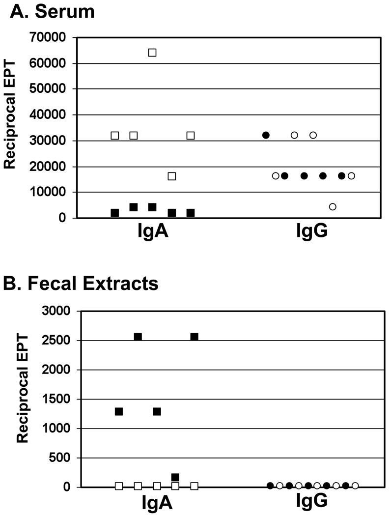
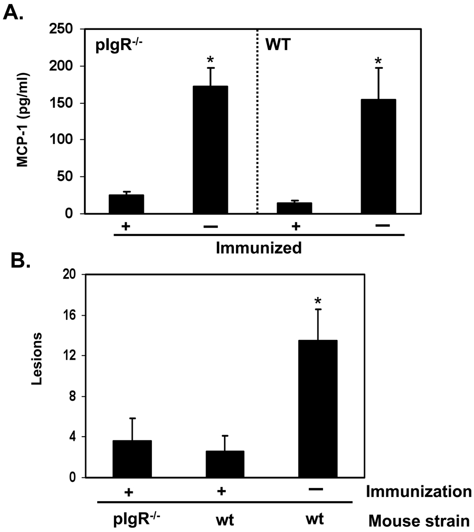
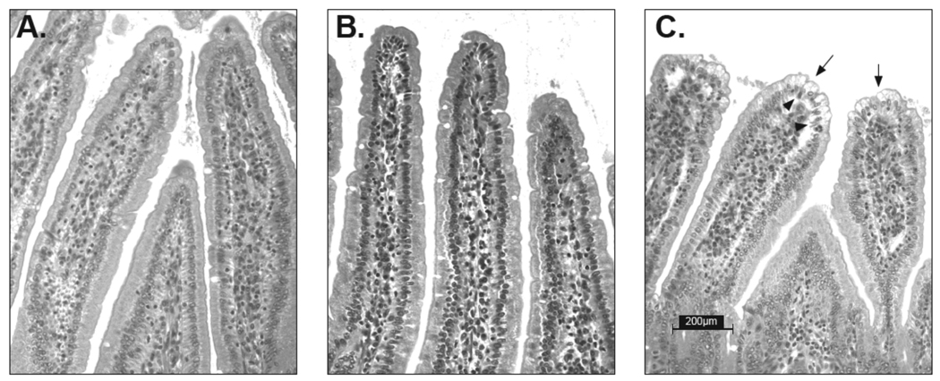
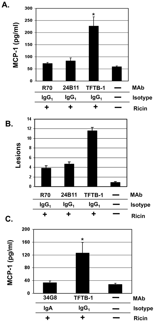
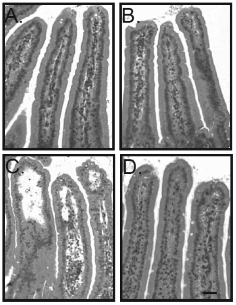
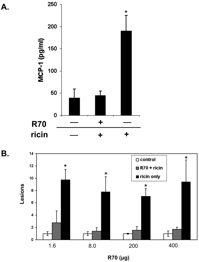
The RNA N-glycosidase ribosome inactivating proteins (RIPs) constitute a ubiquitous family of plant- and bacterium-derived toxins that includes the category B select agents ricin, abrin and shiga toxin. While these toxins are potent inducers of intestinal epithelial cell death and inflammation, very little is known about the mechanisms underlying mucosal immunity to these toxins. In the present study, we report that secretory IgA (SIgA) antibodies are not required for intestinal immunity to ricin, as evidenced by the fact that mice devoid of SIgA, due to a mutation in the polymeric immunoglobulin receptor, were impervious to the effects of intragastric toxin challenge following ricin toxoid immunization. Furthermore, parenteral administration of ricin-specific monoclonal IgGs, directed against either ricin's enzymatic subunit (RTA) or ricin's binding subunit (RTB), to wild type mice was as effective as monoclonal IgAs with comparable specificities in imparting intestinal immunity to ricin. These data are consistent with reports from others demonstrating that immunization of mice by routes known not to induce mucosal antibody responses (e.g., intramuscular and intradermal) is sufficient to elicit protection against both systemic and mucosal ricin challenges.


Figures

Fig. 1
Fig. 1. Anti-toxin serum and fecal antibody titers in wild type and pIgR−/− mice following immunization with ricin toxoid
Groups of wild type (solid symbols) and pIgR−/− (open symbols) mice (n=5–8 mice per group) were immunized 3 times with RT at 10–14 day intervals by the i.g. route. Seven days after the final immunization, anti-toxin IgA and IgG levels were measured in (A) serum and (B) fecal extracts. Each symbol represents an individual mouse. EPT, end point titer.
Fig. 2
Fig. 2. Intestinal immunity to ricin in the absence of SIgA
Groups of wild type and pIgR−/− mice (5–8 mice per group) were immunized (+) or not (−) with RT, as described in the legend of Fig. 1, and then challenged 7–10 days later with ricin (5 mg/kg) by the i.g. route. Intestinal tissues were collected 24 hr later. (A) MCP-1 levels were determined in intestinal homogenates. (B) H&E stained tissue sections were scored for lesions, as described in the Materials and Methods. The asterisks indicate that MCP-1 levels (A) and intestinal lesions (B) in control, toxin-challenged mice were statistically different than RT-immunized mice (p<0.05), as determined by the Student’s t test.
Fig. 3
Fig. 3. Absence of ricin-induced epithelial damage in pIgR knock-out mice
Representative images of H&E stained tissue sections from ricin-challenged control, and RT-immunized wild type and pIgR−/− mice, as described in the legends of Figures 1 and 2. (A) RT-immunized, ricin-challenged wild type Balb/c mice; (B) RT-immunized, ricin-challenged pIgR−/− mice; (C) ricin-challenged, control Balb/c mice. Arrows indicate evidence of epithelial cell vacuolization; arrowheads indicated sites of epithelial detachment. The images were originally captured using a 25 × objective. Scale bar equals 200 µm.
Fig. 4
Fig. 4. Intestinal immunity to ricin following parenteral immunization with RiVax
Groups of Balb/c mice (n= 6–9/group) were immunized by the s.c. route three times with RiVax (10 µg/immunization) or PBS. Serum was collected 7–10 days following each immunization. (A) Anti-ricin serum IgG antibody titers were determined by ELISA, as described in Materials and Methods. Shaded circles reflect immunized mice, whereas open triangles reflect sham-immunized controls. Each symbol represents a single mouse. The solid, horizontal bars reflect the average of the groups. (B) Groups of RiVax or sham immunized mice were challenged with ricin (5 mg/kg) by gavage. A third group of mice not immunized or challenged served as negative controls for this experiments. MCP-1 levels in intestinal tissues from all three groups of mice were determined 24 hr following intragastric ricin challenge, as described in Materials and Methods. The asterisks indicate that MCP-1 levels in control, toxin-challenged mice were statistically different from RiVax-immunized mice (p<0.05), as determined by the Student’s t test.
Fig. 5
Fig. 5. Passively administered toxin-specific IgG MAbs protect the intestinal epithelium from ricin
Groups of female BALB/c mice bearing hybridoma backpacks secreting the indicated IgG or IgA MAbs were challenged i.g. with ricin (5 mg/kg). 24 hr later, the mice were euthanized and intestinal tissues were assessed for MCP-1 (panels A, C) or scored for mucosal lesions (panel B). Each column represents the average values (+/− s.d.) of a single experiment with a total 5–8 mice per group. The asterisks in panels A and C indicate that the mean MCP-1 concentration is significantly greater (p<0.05) than the mean concentration determined from untreated control animals (far right columns in panels A, C).
Fig. 6
Fig. 6. Anti-ricin IgG MAbs protect against toxin-induced epithelial damage
Representative images of intestinal tissues collected 24 hr after mice carrying backpack tumors secreting IgG MAbs (A) R70, (B) 24B11, or (C) TFTB-1 were challenged with ricin. Panel D shows tissues from a control mouse that had not been challenged with ricin. Scale bar equals 200 µm.
Fig. 7
Fig. 7. IgG MAb R70 administered systemically confers intestinal immunity to ricin
MAb R70 (8 µg) was administered by i.p injection to a single group of BALB/c mice. 12 hr later, the R70-treated mice, as well as a control group of mice, were challenged by gavage with ricin (5 mg/kg) diluted into PBS. A third group of animals, not treated with R70, received PBS only by gavage. All three groups of mice were euthanized 18 hr following challenge, and MCP-1 levels were assessed in intestinal homogenates. Each column represents the average (+/− s.d.) of a single experiment, with a total of 6 mice per group. The asterisk indicates that MCP-1 in sham-treated, toxin-challenged mice were statistically different from unchallenged controls (p<0.05), as determined by the Student’s t test. (B) Groups of mice (n=5–6) were administered R70 at the indicated doses by i.p injection then challenged 24 hr later with ricin by gavage. Intestinal tissues were collected 24 hr later and scored for damage. The asterisks indicate lesion scores that were significantly greater (p<0.05) than control, unchallenged mice,
Fig. 8
Fig. 8. IgG-mediated intestinal immunity to ricin is independent of FcRn
MAb R70 (10 µg) was administered by i.p injection to β2M−/− mice or C57Bl/6 age and sex-matched controls. Twenty-four hours later, the mice were challenged i.g. with ricin (5 mg/kg). The animals were then euthanized 24 hr following challenge, and MCP-1 levels were assessed in intestinal homogenates. Each column represents the average values (+/− s.d.) of a single experiment, with a total 6 mice per group. The asterisks indicate that the respective MCP-1 values between groups of identically treated β2M−/− and C57BL/6 mice were not statistically significant.

References

    1. Endo Y, Mitsui K, Motizuki M, Tsurugi K. The mechanism of action of ricin and related toxins on eukaryotic ribosomes. J Biol Chem. 1987;262:5908–5912. - PubMed
    1. Baenziger JU, Fiete D. Structural determinants of Ricinus communis agglutinin and toxin specificity for oligosaccharides. JBiolChem. 1979;254(19):9795–9799. - PubMed
    1. Rutenber E, Ready M, Robertus JD. Structure and evolution of ricin B chain. Nature. 1987;326(6113):624–626. - PubMed
    1. Zentz C, Frenoy JP, Bourrillon R. Binding of galactose and lactose to ricin. Equilibrium studies. Biochim Biophys Acta. 1978 Sep 26;536(1):18–26. - PubMed
    1. Rapak A, Falnes PO, Olsnes S. Retrograde transport of mutant ricin to the endoplasmic reticulum with subsequent translocation to cytosol. Proc Natl Acad Sci U S A. 1997 Apr 15;94(8):3783–3788. - PMC - PubMed

Publication types

MeSH terms