. 2011 Feb 11;286(6):4420-8.
doi: 10.1074/jbc.M110.188730. Epub 2010 Nov 29.

Crystal structures of a cysteine-modified mutant in loop D of acetylcholine-binding protein

Affiliations

Crystal structures of a cysteine-modified mutant in loop D of acetylcholine-binding protein

Marijke Brams et al. J Biol Chem. .

Abstract

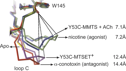
Covalent modification of α7 W55C nicotinic acetylcholine receptors (nAChR) with the cysteine-modifying reagent [2-(trimethylammonium)ethyl] methanethiosulfonate (MTSET(+)) produces receptors that are unresponsive to acetylcholine, whereas methyl methanethiolsulfonate (MMTS) produces enhanced acetylcholine-gated currents. Here, we investigate structural changes that underlie the opposite effects of MTSET(+) and MMTS using acetylcholine-binding protein (AChBP), a homolog of the extracellular domain of the nAChR. Crystal structures of Y53C AChBP show that MTSET(+)-modification stabilizes loop C in an extended conformation that resembles the antagonist-bound state, which parallels our observation that MTSET(+) produces unresponsive W55C nAChRs. The MMTS-modified mutant in complex with acetylcholine is characterized by a contracted C-loop, similar to other agonist-bound complexes. Surprisingly, we find two acetylcholine molecules bound in the ligand-binding site, which might explain the potentiating effect of MMTS modification in W55C nAChRs. Unexpectedly, we observed in the MMTS-Y53C structure that ten phosphate ions arranged in two rings at adjacent sites are bound in the vestibule of AChBP. We mutated homologous residues in the vestibule of α1 GlyR and observed a reduction in the single channel conductance, suggesting a role of this site in ion permeation. Taken together, our results demonstrate that targeted modification of a conserved aromatic residue in loop D is sufficient for a conformational switch of AChBP and that a defined region in the vestibule of the extracellular domain contributes to ion conduction in anion-selective Cys-loop receptors.


Figures

FIGURE 1.
FIGURE 1.
A, left, crystal structure of Aplysia Y53C AChBP modified with MTSET+. The asymmetric unit contains two pentamers, which are in an upside-down orientation relative to each other and interact through two neighboring C-loops (indicated with arrow). The MTSET+-modified side chain is shown as spheres. Right, detailed stereoview of the ligand-binding pocket at the interface of two subunits. The principal face (+) is shown in yellow, complementary face (−) in blue. Conserved aromatic amino acids of the principal face as well as residues involved in interactions with the MTSET+-modified side chain (Gln-36, Tyr-91, and Ser-165) are shown in stick representation. 2FoFc electron density around the Cys-55 side chain is shown as blue mesh with a contour level of 1.0 sigma. B, left, co-crystal structure of Y53C AChBP modified with MMTS and acetylcholine. The asymmetric unit contains 1 pentamer, shown along the 5-fold symmetry axis. The MMTS side chain and acetylcholine molecules are shown in sphere representation. Right, detailed stereoview of the ligand-binding pocket formed by a principal (yellow) and complementary (blue) subunit. Residues involved in ligand-receptor interactions are shown in stick representation. 2FoFc density around the acetylcholine molecules and MMTS-modified side chain is shown as a blue mesh at a contour level of 1.0 sigma. C, sequence alignment showing the conservation of an aromatic amino acid (shown in “black”) in loop D of different Cys-loop receptors. D, stereoview of ligand contacts in the published crystal structure of Lymnaea AChBP in complex with a single carbamylcholine molecule (PDB code 1UV6 and Ref. 20).
FIGURE 2.
FIGURE 2.
Comparison of C-loop conformation for Y53C-MTSET+ (magenta), Y53C-MMTS in complex with acetylcholine (blue), nicotine-bound state (green, PDB code 2BR7, Ref. 20), α-conotoxin ImI-bound state (red, PDB code 2C9T, Ref. 11), and apo state (yellow, PDB code 2W8E, Ref. 21). The dashed lines indicate the distances between the carbonyl oxygen of the conserved Tyr-145 residue in Aplysia AChBP and the γS atom of the vicinal disulfide bond.
FIGURE 3.
FIGURE 3.
A, MMTS-Y53C crystal structure of AChBP shows localized phosphate ions in the vestibule. The pentamer is shown along the 5-fold axis of symmetry with the N terminus pointing away from the viewer. B, detailed view of the anion-binding site in AChBP as seen along the 5-fold symmetry axis from the intracellular side. Ten phosphate ions (yellow) are bound and interact with Lys-40 and Arg-95 (green), Glu-47 and Asp-49 (orange). Phosphate ions and interacting residues are shown in stick representation. The blue mesh around phosphate ions is electron density from a 2FoFc map contoured at a level of 1.0 sigma. Dashed lines indicate distances between two neighboring phosphate ions, 9.3 Å for the upper layer (ring 1), and 7.1 Å for the lower layer (ring 2). C, detailed interaction of residues from a single subunit with phosphate ions in the vestibule. Water molecules are represented as red spheres. Dashed lines indicate hydrogen bonds. D, stereoview from the side of the anion-binding site shows two layers of phosphate ions in a pentagonal arrangement. For clarity, phosphate ions are shown as yellow spheres with a radius of 1 Å. The upper layer (ring 1) matches the position of bound sulfate ions in a previously published study (23).
FIGURE 4.
FIGURE 4.
A, side view of the GLIC pore (8) illustrates the relative position of the homologous ring sites and the selectivity filter at the bottom of transmembrane segments M1 and M2. The sequence alignment shows the conservation of charged residues in ring 1 (Arg-95 in AChBP) and ring 2 (Glu-47 and Asp-49 in AChBP) among different members of the Cys-loop receptor family. B, single channel responses were obtained from wild-type and mutant α1 GlyR using 100 μm glycine in the cell-attached configuration at −30, −60, and −90 mV. Mutant ring 1 contains K104A and G105D, mutant ring 2 contains D57I and R59T, mutant ring 1 + 2 is the quadruple mutant. B, wild-type α1 GlyR; C, mutant ring 1; D, mutant ring 2; E, mutant ring 1 + 2. Mutant ring 2 was only active at −60 and −90 mV. All-point histograms of current amplitude (bin width of 0.1 pA) were fitted by the sum of two Gaussian functions (right side; B, C, D, E). F, single channel conductances were derived from the slope of the current-voltage relationship; (72 pS) wild type, (46 pS) mutant ring 1, (27 pS) mutant ring 2, and (23 pS) mutant ring 1 + 2.

References

    1. Taly A., Corringer P. J., Guedin D., Lestage P., Changeux J. P. (2009) Nat. Rev. Drug Discov. 8, 733–750 - PubMed
    1. Unwin N. (2005) J. Mol. Biol. 346, 967–989 - PubMed
    1. Brejc K., van Dijk W. J., Klaassen R. V., Schuurmans M., van Der Oost J., Smit A. B., Sixma T. K. (2001) Nature 411, 269–276 - PubMed
    1. Smit A. B., Syed N. I., Schaap D., van Minnen J., Klumperman J., Kits K. S., Lodder H., van der Schors R. C., van Elk R., Sorgedrager B., Brejc K., Sixma T. K., Geraerts W. P. (2001) Nature 411, 261–268 - PubMed
    1. Dellisanti C. D., Yao Y., Stroud J. C., Wang Z. Z., Chen L. (2007) Nat. Neurosci. 10, 953–962 - PubMed

Publication types

LinkOut - more resources