. 2011 Feb 4;286(5):3851-62.
doi: 10.1074/jbc.M110.190959. Epub 2010 Nov 29.

Genetic and biochemical analysis of high iron toxicity in yeast: iron toxicity is due to the accumulation of cytosolic iron and occurs under both aerobic and anaerobic conditions

Affiliations

Genetic and biochemical analysis of high iron toxicity in yeast: iron toxicity is due to the accumulation of cytosolic iron and occurs under both aerobic and anaerobic conditions

Huilan Lin et al. J Biol Chem. .

Abstract

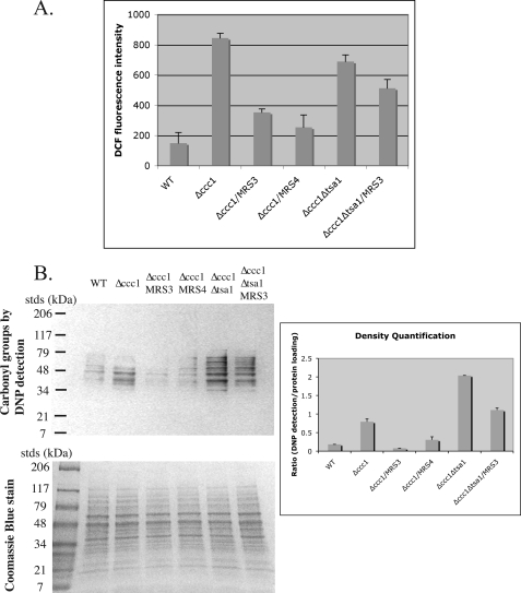
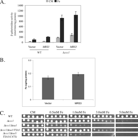
Iron storage in yeast requires the activity of the vacuolar iron transporter Ccc1. Yeast with an intact CCC1 are resistant to iron toxicity, but deletion of CCC1 renders yeast susceptible to iron toxicity. We used genetic and biochemical analysis to identify suppressors of high iron toxicity in Δccc1 cells to probe the mechanism of high iron toxicity. All genes identified as suppressors of high iron toxicity in aerobically grown Δccc1 cells encode organelle iron transporters including mitochondrial iron transporters MRS3, MRS4, and RIM2. Overexpression of MRS3 suppressed high iron toxicity by decreasing cytosolic iron through mitochondrial iron accumulation. Under anaerobic conditions, Δccc1 cells were still sensitive to high iron toxicity, but overexpression of MRS3 did not suppress iron toxicity and did not result in mitochondrial iron accumulation. We conclude that Mrs3/Mrs4 can sequester iron within mitochondria under aerobic conditions but not anaerobic conditions. We show that iron toxicity in Δccc1 cells occurred under both aerobic and anaerobic conditions. Microarray analysis showed no evidence of oxidative damage under anaerobic conditions, suggesting that iron toxicity may not be solely due to oxidative damage. Deletion of TSA1, which encodes a peroxiredoxin, exacerbated iron toxicity in Δccc1 cells under both aerobic and anaerobic conditions, suggesting a unique role for Tsa1 in iron toxicity.


Figures

FIGURE 1.
FIGURE 1.
Expression of Rim2 rescues iron toxicity in Δccc1 cells. Cells deleted for CCC1 were transformed with a plasmid containing a genomic copy of MRS3, CCC1, RIM2, or a control plasmid. The cells were plated on CM with or without iron and grown at 30 °C for 3 days.
FIGURE 2.
FIGURE 2.
Deletion of Rim2 affects mitochondrial functions. A, sporulation of a heterozygous diploid deletion, Δrim2, showed large and small colonies when grown on YPD. B, wild type, wild type rho0, Δrim2, and Δrim2 transformed with a RIM2-expressing plasmid were spotted on glycerol-ethanol-containing (G/E) medium and grown at 30 °C for 3 days. C, wild type rho0, Δmrs3Δmrs4 rho0, Δrim2, or Δmrs3Δmrs4Δrim2 cells were streaked on CM medium or BPS2 and grown for 3 days. D, aconitase activity was assayed in cells from C. Protein concentration was determined, and aconitase activity was normalized to protein concentrations.
FIGURE 3.
FIGURE 3.
Overexpressed Mrs3-FLAG localizes to mitochondria. A, Δccc1 cells were transformed with a control vector or MRS3-FLAG. Cells were incubated in BPS2, CM medium, or CM medium containing 250 μm iron overnight. The levels of Mrs3-FLAG or mitochondrial porin were assayed by Western blot. B, Δccc1 cells transformed with a control vector or MRS3-FLAG were grown in CM medium in the presence orabsence of 250 μm iron overnight. The expression of Mrs3-FLAG and mitochondrial porin wasexamined by immunofluorescence. C, cells transformed with a control plasmid or MRS4-FLAG were grown overnight in CM medium, and the localization of Mrs4-FLAG and mitochondrial porin was examined by immunofluorescence.
FIGURE 4.
FIGURE 4.
Effect of overexpression of Mrs3-FLAG on whole cell and mitochondrial iron. A, wild type or Δccc1 cells transformed with a control vector or MRS3-FLAG were incubated in CM containing 250 μm iron. About 2 × 10⁸ cells were harvested and digested with nitric acid at 80 °C for 1 h. Whole cell iron was determined by an inductively coupled plasma optical emission spectrometer. B, wild type and Δccc1 cells were transformed with a plasmid expressing CCC1-lacZ and either a control vector or MRS3-FLAG. Cells were incubated overnight in CM medium or CM medium with 250 μm iron, and β-galactosidase activity was determined. C, Δccc1 cells were transformed with a control vector or MRS3-FLAG. The cells were incubated in CM medium or CM medium with 50 or 250 μm iron overnight. Mitochondria were isolated, and the amount of mitochondrial iron was determined by an inductively coupled plasma optical emission spectrometer and normalized to mitochondrial protein concentrations. Error bars represent the S.E. of three separate experiments.
FIGURE 5.
FIGURE 5.
Iron induces oxidative damage in Δccc1 cells. A, wild type (DY150), Δccc1, and Δccc1Δtsa1 cells were transformed with empty vector, MRS3-FLAG, or MRS4-FLAG. Cells were incubated with 3 mm iron for 3 h. DCFDA was added after the 2nd h of incubation, and cells were incubated for an additional 1 h. The cells were lysed, and fluorescence was determined. The data were normalized to protein concentration, and error bars represent the S.E. of n = 3 experiments. B, wild type (DY150), Δccc1, and Δccc1Δtsa1 cells were transformed with empty vector, MRS3-FLAG, or MRS4-FLAG. Cells were incubated with 3 mm iron for 3 h. Cells were harvested and homogenized with glass beads. The oxidant modification of proteins was examined by Western blot following derivatization of protein carbonyl groups by 2,4-dinitrophenylhydrazine and incubation with antibodies specific to 2,4-dinitrophenyl (DNP). The upper left panel is an OxyBlot, and the lower left panel is a Coomassie Blue-stained gel.
FIGURE 6.
FIGURE 6.
Deletion of antioxidant genes affects high iron sensitivity. Wild type (DY150), Δccc1, Δyap1, Δtrx2, Δtsa1, and double knock-out strains Δccc1Δyap1, Δccc1Δtsa1, and Δccc1Δtrx2 (A) or Δgrx4 and Δccc1Δgrx4 cells (B) were grown in CM medium. Serial dilutions were spotted onto CM or CM plates containing the specified concentrations of iron. Plates were incubated at 30 °C for 2 days. C, DY150, Δccc1, and Δccc1Δtsa1 cells transformed with empty vector, TSA1, or TSA1(C47S) plasmids were spotted on CM-Ura plates with different concentrations of iron. Plates were incubated at 30 °C for 2 days. D, DY150, Δccc1, Δtrr1, or Δccc1Δtrr1 cells were spotted onto iron plates and grown at 30 °C for 2 days. E, cells as described in D were spotted on plates with addition of H2O2 and grown for 2 days.
FIGURE 7.
FIGURE 7.
Overexpression of Mrs3-FLAG does not protect anaerobically grown Δccc1 cells from high iron toxicity. A, cells transformed with empty vector or MRS3-FLAG were grown aerobically or anaerobically in CM media. Cells were harvested and homogenized with glass beads. Protein levels were analyzed by Western blot using antibodies directed against FLAG and porin. B, wild type, Δmrs3Δmrs4, or Δmrs3Δmrs4 cells transformed with either a control plasmid, MRS3-FLAG, or MRS4-FLAG were grown anaerobically in CM or BPS5 for 3 days. C, Δccc1 cells transformed with empty vector or MRS3-FLAG were grown in CM with 250 μm iron aerobically or anaerobically. Cells were examined by immunofluorescence using antibodies against FLAG and porin. D, wild type cells transformed with empty vector or Δccc1 cells transformed with empty vector, MRS3, ZRC1, or ZRC1(N44I) were spotted onto CM plates or CM plates containing 3 mm iron. Plates were incubated aerobically or anaerobically at 30 °C for 2–3 days.
FIGURE 8.
FIGURE 8.
Overexpression of Mrs3-FLAG anaerobically does not reduce cytosolic iron. A, wild type and Δccc1 cells transformed with empty vector or MRS3-FLAG were also transformed with a CCC1-lacZ plasmid. The cells were grown anaerobically and incubated in the presence or absence of 250 μm iron. Cells were harvested, and β-galactosidase activity was determined. Error bars represent the S.E. from n = 3 experiments. B, mitochondria were isolated from Δccc1 cells transformed with empty vector or MRS3-FLAG grown anaerobically and exposed to 50 μm iron. The amount of iron in mitochondria was determined by an inductively coupled plasma optical emission spectrometer and normalized to mitochondrial protein concentrations. Error bars represent the S.E. from n = 3 experiments. C, wild type (DY150), Δccc1, and Δccc1Δtsa1 cells with empty vector and Δccc1Δtsa1 cells with TSA1 or TSA1(C47S) plasmid were spotted on plates with different concentrations of iron and grown for 3 days.

Similar articles

Cited by

References

    1. Touati D. (2000) Arch. Biochem. Biophys. 373, 1–6 - PubMed
    1. Valko M., Morris H., Cronin M. T. (2005) Curr. Med. Chem. 12, 1161–1208 - PubMed
    1. Pietrangelo A., Trautwein C. (2004) Nat. Clin. Pract. Gastroenterol. Hepatol. 1, 39–45 - PubMed
    1. Philpott C. C., Rashford J., Yamaguchi-Iwai Y., Rouault T. A., Dancis A., Klausner R. D. (1998) EMBO J. 17, 5026–5036 - PMC - PubMed
    1. Milgrom E., Diab H., Middleton F., Kane P. M. (2007) J. Biol. Chem. 282, 7125–7136 - PubMed

Publication types