. 2011 Jan;22(1):43-8.
doi: 10.1681/ASN.2010040373. Epub 2010 Nov 29.

MCP-1 contributes to arteriovenous fistula failure

Affiliations

MCP-1 contributes to arteriovenous fistula failure

Julio P Juncos et al. J Am Soc Nephrol. 2011 Jan.

Abstract

Vascular access dysfunction compromises the care of patients on chronic hemodialysis. Elucidating the mechanisms of such dysfunction and devising strategies that may interrupt neointimal hyperplasia and relevant pathogenetic pathways are essential. Here, we show that, in the venous segment of a murine model of an arteriovenous fistula, monocyte chemoattractant protein-1 (MCP-1) mRNA and protein increase, accompanied by increased activity of the transcription factors NF-κB and AP-1. Genetic deficiency of MCP-1 proved markedly protective in this murine model, reflected by increased fistula patency 6 weeks after its formation, decreased venous wall thickness, and increased luminal area. An early effect of MCP-1 deficiency was the attenuation of the marked induction of CCL5 (RANTES) that occurred in this model, a chemokine recently recognized as a critical participant in vascular injury. Finally, in a rat model of an arteriovenous fistula, we localized expression of MCP-1 to the endothelium, proliferating smooth muscle cells and infiltrating leukocytes. In summary, marked upregulation of MCP-1 occurs in the venous segment of an arteriovenous fistula in rodents, and this vasculopathic chemokine contributes to failure of the fistula.


Figures
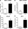
Figure 1.
Figure 1.
Expression of MCP-1 and relevant MCP-1 transcription factors 1 week after establishing an AVF in mice. In the venous segment of the AVF and in the contralateral, nonoperated, jugular vein (Control), measurements of (A) MCP-1 mRNA (Control: n = 7; AVF: n = 9), (B) MCP-1 protein (Control: n = 4; AVF: n = 8), (C) NF-κB (n = 6 in both groups), and (D) AP-1 (n = 6 in both groups) were undertaken. MCP-1 mRNA expression was determined by quantitative real-time reverse transcriptase-PCR, MCP-1 protein expression by ELISA, NF-κB activation by a chemiluminescence-based assay kit, and AP-1 activation by a colorimetric assay kit. For MCP-1 protein analysis by ELISA, four control veins and two AVF veins were pooled for each determination. For all other analyses, one vein was used for each determination.
Figure 2.
Figure 2.
Patency, morphometry, and histology of the AVF in MCP-1+/+ and MCP-1−/− mice 6 weeks after formation of the AVF. (A) The patency of the AVF (left), venous wall thickness (middle), and luminal area/total cross-sectional area (right) 6 weeks after establishing the AVF in MCP-1+/+ and MCP-1−/− mice. In MCP-1+/+ mice, n = 12 to 13, whereas in MCP-1−/− mice, n = 11 to 12. (B) Representative sections of the venous wall in the AVF in MCP-1+/+ mice at magnifications of 200× (top left panel) and 400× (top right panel) and in MCP-1−/− mice at magnifications of 200× (bottom left panel) and 400× (bottom right panel). The venous wall of the AVF in MCP-1+/+ mice compared with the AVF in MCP-1−/− mice is increased in thickness because of the presence of cellular infiltration and increased matrix (neointimal hyperplasia [NH]); additionally, there is organized thrombus (T) adherent to the venous wall of the AVF in MCP-1+/+ mice, and fresh clot (C) in the lumen. All sections were stained with hematoxylin and eosin.
Figure 3.
Figure 3.
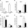
Expression of genes in the venous segment of the AVF 1 week after the formation of an AVF in MCP-1+/+ and MCP-1−/− mice. Real-time RT-PCR was used to determine mRNA expression in the venous segment of the AVF and in the contralateral, nonoperated, jugular vein (Control) of MCP-1+/+ (open bars) and MCP-1−/− (closed bars) mice. Shown are the mRNA expression levels of MCP-1 (A), HO-1 (B), BMP-7 (C), and CCL5 (RANTES) (D). In MCP-1+/+ mice, n = 7 for Control and n = 8 for AVF measurements, whereas in MCP-1−/− mice, n = 8 for both Control and AVF measurements. *P < 0.05 for CCL5 mRNA expression in the venous segment in the AVF in MCP-1−/− versus MCP-1+/+ mice. ND, MCP-1 mRNA was not detectable in the venous segments of the Control or AVF veins in MCP-1−/− mice.
Figure 4.
Figure 4.
MCP-1 expression by immunohistochemistry in the femoral AVF in the rat 5 weeks after the formation of the AVF. Immunohistochemistry studies were performed to localize expression of MCP-1 in the venous segment of the femoral AVF in the rat 5 weeks after establishing the AVF. Studies were performed with nonimmune serum for the sham-operated vein (A) and the venous segment of the AVF (B) and with an MCP-1 antibody for the sham-operated vein (C) and the venous segment of the AVF (D). MCP-1 is detected in endothelial cells (E), smooth muscle cells (SM), and leukocytes (L) in the AVF, whereas endothelial cells express MCP-1 in the sham-operated vein. All sections are shown at a magnification of 400×.

References

    1. Agarwal A, Segal MS: Intimal exuberance: Veins in jeopardy. Am J Pathol 162: 1759–1761, 2003 - PMC - PubMed
    1. Allon M: Current management of vascular access. Clin J Am Soc Nephrol 2: 786–800, 2007 - PubMed
    1. Durante W, Lin CC: Homing in on arteriovenous fistula survival. Kidney Int 74: 9–11, 2008 - PMC - PubMed
    1. Roy-Chaudhury P, Spergel LM, Besarab A, Asif A, Ravani P: Biology of arteriovenous fistula failure. J Nephrol 20: 150–163, 2007 - PubMed
    1. Schwab SJ, Harrington JT, Singh A, Roher R, Shohaib SA, Perrone RD, Meyer K, Beasley D: Vascular access for hemodialysis. Kidney Int 55: 2078–2090, 1999 - PubMed

Publication types