Review
. 2011 Feb 1;589(Pt 3):481-8.
doi: 10.1113/jphysiol.2010.199539. Epub 2010 Nov 29.

Visualization of glutamate as a volume transmitter

Affiliations
Review

Visualization of glutamate as a volume transmitter

Yohei Okubo et al. J Physiol. .

Abstract

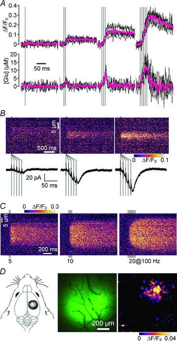
Glutamate is the major excitatory neurotransmitter in the central nervous system. Although glutamate mediates synaptically confined point-to-point transmission, it has been suggested that under certain conditions glutamate may escape from the synaptic cleft (glutamate spillover), accumulate in the extrasynaptic space, and mediate volume transmission to regulate important brain functions. However, the inability to directly measure glutamate dynamics around active synapses has limited our understanding of glutamatergic volume transmission. The recent development of a family of fluorescent glutamate indicators has enabled the visualization of extrasynaptic glutamate dynamics in brain tissues. In this topical review, we examine glutamate as a volume transmitter based on novel results of glutamate imaging in the brain.


Figures

Figure 1
Figure 1. Neural and glial functions mediated by glutamatergic volume transmission
Left, neuronal functions mediated by glutamatergic volume transmission. AMPAR, NMDAR, kainate receptor and mGluR localised to various extrasynaptic locations or within neighbouring synapses are activated by glutamate spillover to mediate physiological and pathophysiological events. Right, astrocytic functions mediated by glutamatergic volume transmission. Astrocytic mGluR is activated by glutamate spillover to induce gliotransmitter release and haemodynamic response.
Figure 2
Figure 2. Imaging of extrasynaptic glutamate dynamics by EOS
A, a schematic of the EOS structure. EOS is composed of the recombinant S1S2 domain of GluR2 (in magenta) and a conjugated fluorescent dye (in green). B, dose–response relationship between glutamate concentration and fractional change in fluorescence intensity (ΔF/F0) of EOS sister indicators. Estimated Kd values of K716A-EOS and L401C-EOS were 174 nm and 1.57 μm, respectively. C, EOS immobilised in the molecular layer of cerebellar slices. The magnified image (lower panel) shows a roughly even distribution of EOS in the extracellular space with silhouettes of cellular components. D, imaging of EOS signals in response to PF stimulation (5 pulses at 100 Hz). ΔF/F0 is colour coded as indicated. Modified from Okubo et al. (2010), copyright (2010) National Academy of Sciences, USA.
Figure 3
Figure 3. Direct evaluation of extrasynaptic glutamate dynamics by EOS imaging
A, temporal summation in extrasynaptic glutamate dynamics upon repetitive PF stimulation in the cerebellar cortex and estimation of corresponding glutamate concentration. Upper traces show the time course of the EOS signal and the lower traces show the corresponding time course of glutamate concentration estimated by the deconvolution method (Okubo et al. 2010). Magenta traces indicate averaged data. B, spatial summation in extrasynaptic glutamate dynamics. The simultaneous measurement of EOS signals and EPSCs upon PF stimulation (5 pulses at 100 Hz) shows a correlation between extrasynaptic glutamate dynamics and PF input density. C, EOS signal upon 5, 10 and 20 pulses of PF stimulation at 100 Hz. Increasing the number of stimulation pulses increased the spatial width of the EOS signal. Peripheral signals emerged after 10 and 20 pulses of stimulation that showed significant latency. D, extrasynaptic glutamate dynamics upon sensory input from the hind paw. EOS was immobilised in the rat sensory cortex corresponding to the hind paw (left and middle panels). Peak EOS signal induced by 200 ms tactile stimulation to the hind paw is shown (right panel). Modified from Okubo et al. (2010), copyright (2010) National Academy of Sciences, USA.

References

    1. Agulhon C, Fiacco TA, McCarthy KD. Hippocampal short- and long-term plasticity are not modulated by astrocyte Ca2+ signaling. Science. 2010;327:1250–1254. - PubMed
    1. Arnth-Jensen N, Jabaudon D, Scanziani M. Cooperation between independent hippocampal synapses is controlled by glutamate uptake. Nat Neurosci. 2002;5:325–331. - PubMed
    1. Asztely F, Erdemli G, Kullmann DM. Extrasynaptic glutamate spillover in the hippocampus: dependence on temperature and the role of active glutamate uptake. Neuron. 1997;18:281–293. - PubMed
    1. Baker DA, Xi ZX, Shen H, Swanson CJ, Kalivas PW. The origin and neuronal function of in vivo nonsynaptic glutamate. J Neurosci. 2002;22:9134–9141. - PMC - PubMed
    1. Barbour B. An evaluation of synapse independence. J Neurosci. 2001;21:7969–7984. - PMC - PubMed

Publication types

MeSH terms

LinkOut - more resources