. 2011 Feb;79(2):695-707.
doi: 10.1128/IAI.01071-10. Epub 2010 Nov 29.

Comparative kinetics of Escherichia coli- and Staphylococcus aureus-specific activation of key immune pathways in mammary epithelial cells demonstrates that S. aureus elicits a delayed response dominated by interleukin-6 (IL-6) but not by IL-1A or tumor necrosis factor alpha

Affiliations

Comparative kinetics of Escherichia coli- and Staphylococcus aureus-specific activation of key immune pathways in mammary epithelial cells demonstrates that S. aureus elicits a delayed response dominated by interleukin-6 (IL-6) but not by IL-1A or tumor necrosis factor alpha

Juliane Günther et al. Infect Immun. 2011 Feb.

Abstract

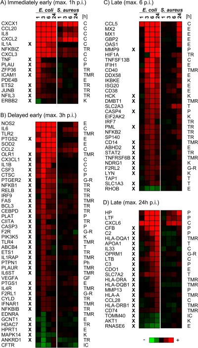
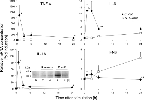
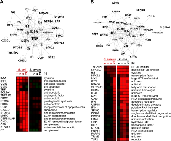
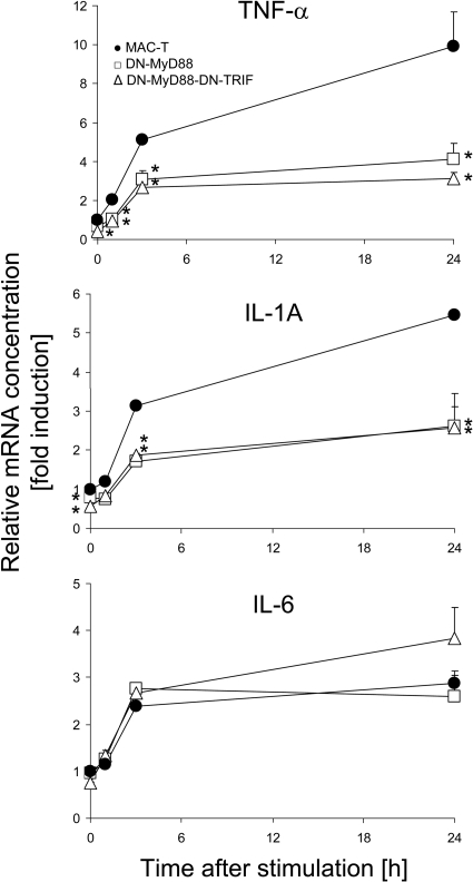
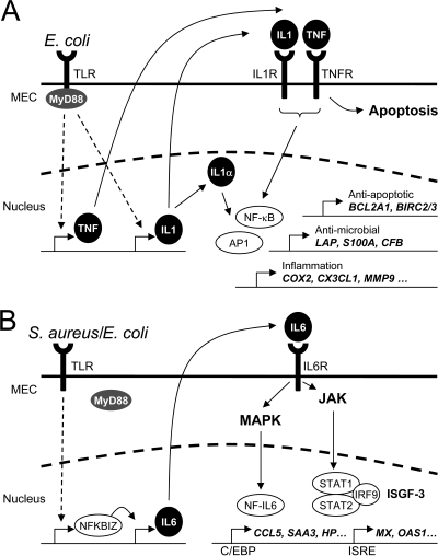
Infections of the udder by Escherichia coli very often elicit acute inflammation, while Staphylococcus aureus infections tend to cause mild, subclinical inflammation and persistent infections. The molecular causes underlying the different disease patterns are poorly understood. We therefore profiled the kinetics and extents of global changes in the transcriptome of primary bovine mammary epithelial cells (MEC) after challenging them with heat-inactivated preparations of E. coli or S. aureus pathogens. E. coli swiftly and strongly induced an expression of cytokines and bactericidal factors. S. aureus elicited a retarded response and failed to quickly induce an expression of bactericidal factors. Both pathogens induced similar patterns of chemokines for cell recruitment into the udder, but E. coli stimulated their synthesis much faster and stronger. The genes that are exclusively and most strongly upregulated by E. coli may be clustered into a regulatory network with tumor necrosis factor alpha (TNF-α) and interleukin-1 (IL-1) in a central position. In contrast, the expression of these master cytokines is barely regulated by S. aureus. Both pathogens quickly trigger an enhanced expression of IL-6. This is still possible after completely abrogating MyD88-dependent Toll-like receptor (TLR) signaling in MEC. The E. coli-specific strong induction of TNF-α and IL-1 expression may be causative for the severe inflammatory symptoms of animals suffering from E. coli mastitis, while the avoidance to quickly induce the synthesis of bactericidal factors may support the persistent survival of S. aureus within the udder. We suggest that S. aureus subverts the MyD88-dependent activation of immune gene expression in MEC.


Figures

FIG. 1.
FIG. 1.
Comparison of differentially expressed genes in response to challenges with E. coli or S. aureus particles. Considered are genes contributing to the group of “inflammatory response” genes, as defined by IPA software. Heat maps (red, increase; green, decrease) visualize changes in mRNA concentrations relative to the unstimulated controls at different times after challenge (p.i.). Genes are grouped according to the time to reach their maximally altered mRNA concentrations after an E. coli challenge. The X symbol indicates genes regulated solely by E. coli. Gene functions according to the IPA annotation are given to the right (C, cytokine; E, enzyme; G-R, G-protein-coupled receptor; IC, ion channel; K, kinase; P, peptidase; Ph, phosphatase; T, transporter; TMR, transmembrane receptor; TR, transporter).
FIG. 2.
FIG. 2.
Alteration in the mRNA concentrations of TNF-α, IL-1A, IL-6, and IFN-β (IFN-β1 and IFN-β2) in pbMEC after challenge with E. coli (⧫) and S. aureus (□) particles. Shown are values for mean fold induction (ordinate, ± standard error of the mean [SEM]; n = 3) at times after challenge (abscissa) relative to the mRNA concentration measured for unstimulated cells (set as 1). Asterisks indicate a statistical significance (*, P ≤ 0.05; **, P ≤ 0.01 [by t test]) of the difference between the E. coli and S. aureus values at the time indicated. The IL-1A insert shows a Western blot analysis of IL-1A in lysates (20 μg/slot) from unchallenged pbMEC (0 h) and from those challenged for 2 h or 4 h with S. aureus or E. coli particles (107 particles/ml). The band shows the 31-kDa IL-1A precursor protein.
FIG. 3.
FIG. 3.
Network analysis of regulatory relationships. (A) E. coli network. IPA analysis reveals an interaction network dominated by IL-1A and TNF-α as having the highest statistical probability (network score of 49) of the 27 top-ranked genes regulated exclusively by E. coli (top). Heat maps (bottom) (red, increase; green, decrease) compare the regulation intensities of those genes after E. coli (right) or S. aureus (left) challenge. (B) Consideration of all S. aureus-regulated gene (n = 138) yields with the highest statistical significance (network score of 48) for a network dominated by IL-6. Heat maps (bottom) show the regulation intensities of those genes after E. coli (right) or S. aureus (left) challenge. ECM, extracellular matrix.
FIG. 4.
FIG. 4.
Impact of functional knockdown of TLR signaling on induced cytokine gene expression. Shown are data for the alteration of TNF-α, IL-1A, and IL-6 mRNA concentrations in WT MAC-T cells or those stably transfected with vectors expressing DN-MyD88 and DN-MyD88-DN-TRIF mutants after challenge with heat-inactivated E. coli particles. Mean fold induction values (ordinate, ±SEM) relative to the mRNA concentrations measured in unstimulated MAC-T cells (set as 1) were derived from one experiment representative of three, which was assayed in triplicate. Asterisks indicate that the mRNA concentrations measured for the DN mutant transfectants are statistically different (*, P ≤ 0.05 [by t test]) from those for WT MAC-T cells at the respective time point after E. coli challenge.
FIG. 5.
FIG. 5.
Proposed signaling pathways being activated in MEC through challenges with heat-inactivated particles of E. coli or S. aureus pathogens. (A) E. coli immediately induces the early expression of the proinflammatory master cytokines IL-1α and IL-1β as well as TNF-α. The activation of these cytokines is MyD88 dependent. IL-1β and TNF-α are synthesized and secreted and may bind to their cognate receptors (IL1R and TNFR). This subsequently leads to the activation of NF-κB and AP1, which in turn induces the expression of the secondary response genes (e.g., BCL2A1, BIRC1/2, LAP, S100A8/9, CFB, COX2, CX3CL1, and MMP9). In addition, TNF-α is also one major mediator of apoptosis. Intracellular IL-1α may also function as an endogenous transcription regulator inside the cell of its synthesis (22). (B) S. aureus and E. coli stimulations both lead to an early activation of NFKBIZ. This transcriptional activator is known to be essential for the TLR-mediated induction of IL-6 expression. We assume that the induction of IL-6 expression in MEC is MyD88 independent. Secreted IL-6 binds to the IL-6 receptor (IL6R), resulting in the activation of both JAK and MAPK signaling cascades. The activation of the JAK cascade leads to the formation of an active ISGF3 complex, which may induce the expression of genes harboring ISRE sites in their promoters. MAPKs activated through IL-6 signaling induce the activation of NF-IL-6, also known as the transcription factor C/EBPβ. The transcription induced by ISGF3 and C/EBPβ dominates the secondary response to S. aureus.

References

    1. Bannerman, D. D. 2009. Pathogen-dependent induction of cytokines and other soluble inflammatory mediators during intramammary infection of dairy cows. J. Anim. Sci. 87:10-25. - PubMed
    1. Bannerman, D. D., et al. 2004. Innate immune response to intramammary infection with Serratia marcescens and Streptococcus uberis. Vet. Res. 35:681-700. - PubMed
    1. Bannerman, D. D., M. J. Paape, W. R. Hare, and J. C. Hope. 2004. Characterization of the bovine innate immune response to intramammary infection with Klebsiella pneumoniae. J. Dairy Sci. 87:2420-2432. - PubMed
    1. Bannerman, D. D., et al. 2004. Escherichia coli and Staphylococcus aureus elicit differential innate immune responses following intramammary infection. Clin. Diagn. Lab. Immunol. 11:463-472. - PMC - PubMed
    1. Bannerman, D. D., M. J. Paape, and A. Chockalingam. 2006. Staphylococcus aureus intramammary infection elicits increased production of transforming growth factor-α, β1, and β2. Vet. Immunol. Immunopathol. 112:309-315. - PubMed

Publication types

MeSH terms

Associated data