. 2010 Dec;8(12):1610-8.
doi: 10.1158/1541-7786.MCR-10-0398. Epub 2010 Nov 29.

Enhancer of zeste homolog 2 promotes the proliferation and invasion of epithelial ovarian cancer cells

Affiliations

Enhancer of zeste homolog 2 promotes the proliferation and invasion of epithelial ovarian cancer cells

Hua Li et al. Mol Cancer Res. 2010 Dec.

Abstract

Enhancer of zeste homolog 2 (EZH2) is the catalytic subunit of the polycomb repressive complex 2 (PRC2) that includes noncatalytic subunits suppressor of zeste 12 (SUZ12) and embryonic ectoderm development (EED). When present in PRC2, EZH2 catalyzes trimethylation on lysine 27 residue of histone H3 (H3K27Me3), resulting in epigenetic silencing of gene expression. Here, we investigated the expression and function of EZH2 in epithelial ovarian cancer (EOC). When compared with primary human ovarian surface epithelial (pHOSE) cells, EZH2, SUZ12, and EED were expressed at higher levels in all 8 human EOC cell lines tested. Consistently, H3K27Me3 was also overexpressed in human EOC cell lines compared with pHOSE cells. EZH2 was significantly overexpressed in primary human EOCs (n = 134) when compared with normal ovarian surface epithelium (n = 46; P < 0.001). EZH2 expression positively correlated with expression of Ki67 (P < 0.001; a marker of cell proliferation) and tumor grade (P = 0.034) but not tumor stage (P = 0.908) in EOC. There was no correlation of EZH2 expression with overall (P = 0.3) or disease-free survival (P = 0.2) in high-grade serous histotype EOC patients (n = 98). Knockdown of EZH2 expression reduced the level of H3K27Me3 and suppressed the growth of human EOC cells both in vitro and in vivo in xenograft models. EZH2 knockdown induced apoptosis of human EOC cells. Finally, we showed that EZH2 knockdown suppressed the invasion of human EOC cells. Together, these data demonstrate that EZH2 is frequently overexpressed in human EOC cells and its overexpression promotes the proliferation and invasion of human EOC cells, suggesting that EZH2 is a potential target for developing EOC therapeutics.


Conflict of interest statement

Disclosure of potential conflicts of interest

No potential conflicts of interest were declared.

Figures
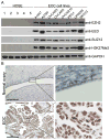
Figure 1
Figure 1. EZH2 is expressed at higher levels in human EOC cell lines and primary human EOCs compared to normal ovarian surface epithelium
(A) Expression of EZH2, EED, SUZ12, H3K27Me3 and GAPDH in five individual batches of pHOSE cell cultures and indicated human EOC cell lines was determined by western blotting. (B) Examples of EZH2 immunohistochemical staining in normal ovary and primary human EOC (shown is an example of high-grade serous histotype EOC). Bar = 50μm.
Figure 2
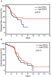
Figure 2. EZH2 expression does not correlate with disease-free or overall survival in high-grade serous histotype EOC patients
The univariate disease-free (A) and overall survival (B) curves (Kaplan-Meier method) for high-grade serous histotype EOC patients with low or high EZH2 protein levels as assessed by immunohistochemistry analysis.
Figure 3
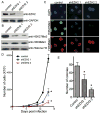
Figure 3. EZH2 knockdown inhibits the growth of SKOV3 cells in vitro
(A) SKOV3 cells were infected with two individual lentivirus encoded shEZH2 or control. Drug-selected cells were examined for expression of EZH2 and GAPDH by western blotting. (B) Same as (A), but examined for expression of H3K27Me3 and H3K9Me3 by western blotting. (C) Same as (A), but examined for expression of H3K27Me3 and H3K9Me3 by immunofluorescence staining. DAPI counter-staining was used to visualize the cell nuclei. (D) Same as (A), but equal number of drug-selected cells were seeded and counted at indicated time points. Mean of three independent experiments with SD. * p<0.001. (E) Same as (A), 1×10⁴ cells were seeded in soft-agar and the number of colonies were counted after three weeks of culture. Mean of three independent experiments with SD. *p<0.001.
Figure 4
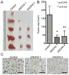
Figure 4. EZH2 knockdown suppresses the growth of SKOV3 cells in vivo in immunocompromised mice
(A) 5×10⁶ control or shEZH2 expressing SKOV3 cells were injected s.c. into immunocompromised nude mice (n=5). Four weeks post injection, tumors were removed from mice. (B) Quantitation of (A), the size of tumors were measured. Mean of tumor sizes with SEM. (C) Xenografted tumors formed by control or shEZH2 expressing SKOV3 cells were sectioned and stained for EZH2 expression. Bar = 50μm.
Figure 5
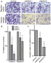
Figure 5. EZH2 knockdown suppresses the invasion of SKOV3 cells
(A) Equal number of control and shEZH2 expressing SKOV3 cells were assayed for migration through uncoated control membrane or invasion through matrigel-coated membrane. The cells migrated through control membrane or invaded through matrigel-coated membrane were stained with 1% crystal violet in PBS. (B) Quantitation of (A). Relative percentage of shEZH2 expressing cells migrated through control membrane or invaded through matrigel-coated membrane compared to controls was indicated. Mean of three independent experiments with SD. *p<0.05. (C) Invasion index of shEZH2 expressing SKOV3 cells compared to controls. Invasion index is the ratio between cells invaded through matrigel-coated membrane and cells migrated through control membrane. Mean of three independent experiments with SD. *p<0.05.
Figure 6
Figure 6. EZH2 knockdown induces apoptosis of SKOV3 cells
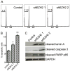
(A) Control and shEZH2 expressing SKOV3 cells were examined for cell cycle distribution by FACS. The percentage of sub-G1 cells was indicated. (B) Control and shEZH2 expressing cells were stained for Annexin V, a cell surface marker of apoptosis. Annexin V positive cells were measured by Guava assay. Mean of three independent experiments with SD. *p=0.019 and ** p<0.001 compared to controls. (C) Same as (B), but examined for expression of cleaved Lamin A, PARP p85 and caspase 3, all markers of apoptosis in control and shEZH2 expressing cells.

References

    1. Cao R, Wang L, Wang H, Xia L, Erdjument-Bromage H, Tempst P, et al. Role of histone H3 lysine 27 methylation in Polycomb-group silencing. Science. 2002;298:1039–43. - PubMed
    1. Czermin B, Melfi R, McCabe D, Seitz V, Imhof A, Pirrotta V. Drosophila enhancer of Zeste/ESC complexes have a histone H3 methyltransferase activity that marks chromosomal Polycomb sites. Cell. 2002;111:185–96. - PubMed
    1. Kuzmichev A, Nishioka K, Erdjument-Bromage H, Tempst P, Reinberg D. Histone methyltransferase activity associated with a human multiprotein complex containing the Enhancer of Zeste protein. Genes Dev. 2002;16:2893–905. - PMC - PubMed
    1. Muller J, Hart CM, Francis NJ, Vargas ML, Sengupta A, Wild B, et al. Histone methyltransferase activity of a Drosophila Polycomb group repressor complex. Cell. 2002;111:197–208. - PubMed
    1. Cao R, Zhang Y. SUZ12 is required for both the histone methyltransferase activity and the silencing function of the EED-EZH2 complex. Mol Cell. 2004;15:57–67. - PubMed

MeSH terms